






Yes, you can tear your ACL without knowing it
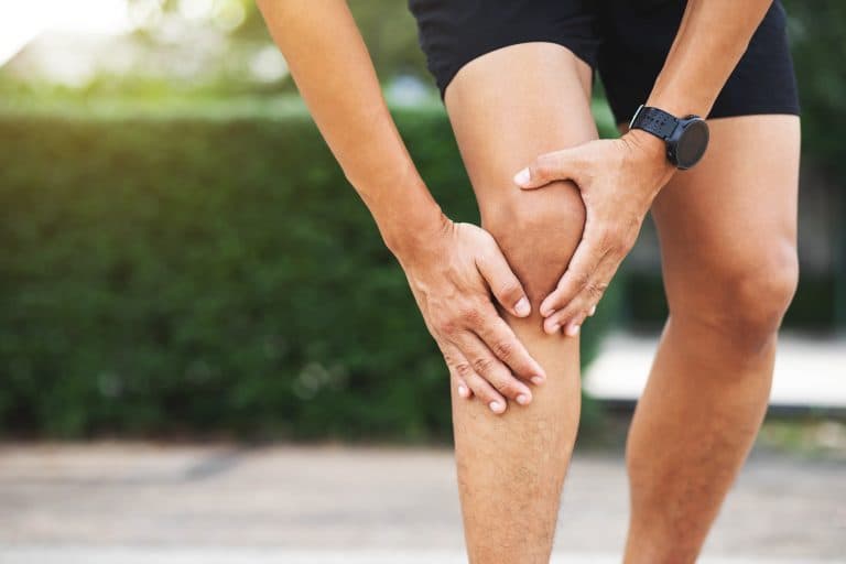
A twist during a weekend five-a-side match, a stumble on a trail run, a clumsy landing from a jump — and then the knee aches for a few days before settling. It feels like a mild sprain, and within a week you are walking normally. For a meaningful number of people, that unremarkable episode is the moment the ACL tore.
The signature triad — an audible pop at the time of injury, rapid swelling within six to twenty-four hours, and a sensation that the knee is giving way — appears in textbook descriptions of ACL rupture, but none of the three is universal. Partial tears, in particular, may produce only brief local pain that resolves quickly, with little or no visible swelling. Even a complete, grade-3 rupture leaves most people able to walk in a straight line, because straight-line gait places relatively modest rotational demand on the joint. Add strong quadriceps and hamstrings to the picture, and the muscular support can compensate for the lost ligament well enough to mask instability during everyday, low-demand movement.
This functional reserve is precisely what allows the injury to go unrecognised. How the knee feels on a short walk is not a reliable guide to whether the ACL is intact. Clinical examination — particularly the Lachman test — combined with MRI is the only way to confirm or exclude the diagnosis.
Why partial tears and mild symptoms are so easy to dismiss
ACL injuries exist on a spectrum — and where a patient sits on that spectrum shapes how dramatic, or how unremarkable, the initial event feels.
At one end sit grade-1 injuries, where ligament fibres are stretched but remain intact; grade-2 represents a partial rupture; grade-3 denotes complete disruption. Grade-1 and grade-2 tears may produce only localised tenderness and a modest degree of puffiness rather than the rapid, balloon-like effusion that accompanies a full haemarthrosis. When swelling takes a day or two to appear — and fades within a week — it is easy to attribute to a routine knock, particularly if the knee remains functional enough for daily tasks.
That functional capacity has a mechanical explanation. During walking, cycling, or straight-line jogging, the rotational and shear forces across the tibiofemoral joint are relatively low; the quadriceps and hamstrings can shoulder much of the load the ACL would ordinarily provide. The problem surfaces when the joint is asked to perform what it was structurally designed to withstand: rapid pivots, sudden deceleration, and lateral cuts — the staple movements of football, netball, and skiing. In these moments, muscular compensation cannot respond quickly enough, and the knee shifts or buckles.
For many patients, it is precisely these episodes of giving way, recurring weeks or months after the original incident, that finally prompt a medical review. By that point, repeated abnormal loading may already have begun its quiet work on the cartilage and meniscus.
The damage that begins at the moment of injury
The harm to cartilage does not wait for instability to develop. At the moment of rupture, the forces involved are substantial enough to compress and shear the joint surfaces directly — and in roughly half of all ACL injuries, articular cartilage or the meniscus sustains damage at that same instant, before any swelling has formed or any instability episode has occurred.
Within hours, blood from ruptured ligament vessels floods the joint, producing what clinicians call a haemarthrosis. This is not merely uncomfortable: the blood carries pro-inflammatory signalling molecules that set off an acute cascade across the entire joint environment. Part of that cascade involves the release of enzymes — including matrix metalloproteinases — that actively break down the structural proteins of cartilage matrix. The knee is not a passive bystander to its own injury; in the acute phase it becomes a chemically hostile environment for the tissue it most needs to protect.
Articular cartilage has almost no blood supply and a negligible capacity for self-repair. Damage initiated in those first hours does not reverse as the swelling settles and the pain fades. A knee that feels broadly normal after ten days may have already sustained changes to its joint surface that are, at best, very slow to stabilise and, at worst, permanent.
A single episode — even one that appears to resolve — is therefore clinically worth assessing promptly. The absence of ongoing symptoms is not evidence that the joint has healed; it may simply mean the acute phase has passed.
How instability keeps grinding cartilage away
Once the acute phase settles, a different and more protracted problem takes over. The ACL ordinarily prevents the tibia from sliding forward on the femur and controls rotational movement under load. Without it, every weighted step, staircase descent, or change of direction allows a degree of abnormal tibial translation and rotation that an intact ligament would suppress within milliseconds.
Think of a car whose wheel tracking is slightly off: the tyre wears unevenly not because of any single dramatic event, but because each revolution compounds a small, consistent misalignment. The knee behaves similarly. The femoral condyles grind against the menisci and the articular cartilage surface during movements the patient may consider entirely ordinary — it is the repetition, rather than any single episode, that accumulates.
The menisci are central to this cascade. Acting as load-distributing shock absorbers between the femur and tibia, they redirect compressive forces across a wider surface area. As chronic instability drives progressive meniscal wear or secondary tearing, that buffering effect diminishes — and the articular cartilage begins to absorb forces it was not designed to bear directly.
Dramatic giving-way events accelerate this process, but they are not its only driver. Low-level micro-instability during activities the patient considers manageable contributes to the same degenerative pattern. Because cartilage has no meaningful blood supply, what is lost incrementally does not return.
The long-term numbers: osteoarthritis risk after ACL injury
All of this matters because the long-term numbers are striking. The reported incidence of posttraumatic osteoarthritis (PTOA) following ACL injury ranges from 18% to 71% across cohort studies, depending on follow-up length and how OA is defined — whether by imaging changes or by symptoms. At the upper end, a widely cited 2013 review (Friel, PMC, 415 citations) places the figure as high as 87% in long-term follow-up populations. The spread is wide because the studies are genuinely heterogeneous; the honest reading is that meaningful OA risk exists across the full range, not only at the top figure.
What is consistent across studies is the timing. Patients with ACL injuries tend to develop knee OA a decade or more earlier than age-matched peers — commonly in their 40s or 50s, well before the age at which the general population typically encounters joint-surface degeneration. Lifetime risk of requiring knee replacement has been estimated at up to seven times that of people with no knee injury history. One mechanistic framing, cited in the same 2013 review, describes an ACL rupture as effectively 'ageing the knee by 30 years'.
Perhaps the most counterintuitive finding for patients considering surgery is this: ACL reconstruction restores mechanical stability and allows return to sport, but has not been shown to prevent the eventual development of OA. Some studies report similar or even elevated OA rates in surgically reconstructed knees compared with conservatively managed ones. This does not make surgery pointless — stability and function are real and meaningful gains — but it underlines that damage already initiated at the moment of injury and during any intervening period of instability cannot be fully reversed by later reconstruction. The case for early recognition rests precisely on limiting how much of that damage accumulates before the problem is identified.
When to get assessed and what that involves
Several clinical signs warrant specialist review beyond an initial GP appointment: a pop heard or felt at the moment of injury, swelling that developed within the first 24 hours, recurrent episodes of the knee giving way during everyday activity, pain or stiffness that persists beyond a few weeks, or a failure to return to pre-injury activity levels. Any single one of these, following a twisting or hyperextension episode, is sufficient reason to seek further assessment rather than waiting.
A specialist consultation typically opens with a structured account of the injury and its aftermath before moving to clinical examination. The Lachman test and the pivot-shift test apply controlled anterior and rotational loads to assess how much the tibia translates under examiner pressure — together they provide substantial diagnostic information before any imaging is obtained. MRI is commonly requested to characterise the extent of ligament disruption and, importantly, to identify concurrent meniscal or chondral injury. A scan that appears normal does not fully exclude a partial tear where the clinical history and examination findings remain suggestive.
The management decision — structured physiotherapy-led rehabilitation or surgical reconstruction — is not predetermined. Age, activity demands, the degree of functional instability, and the presence of associated injuries all shape what is appropriate; the evidence supports a legitimate role for both pathways in different patients, and neither should be assumed without specialist input.
Where imaging reveals concurrent cartilage or meniscal changes, early joint-preservation planning becomes part of that conversation rather than a later afterthought. It is this integration — assessing the ligament, the menisci, and the articular cartilage together and planning accordingly — that characterises the kind of specialist evaluation available at the London Cartilage Clinic on Harley Street; further information and appointments can be found at londoncartilage.com.
- [1] Anterior cruciate ligament injury – Wikipedia. https://en.wikipedia.org/?curid=5811552 https://en.wikipedia.org/?curid=5811552
Frequently Asked Questions
- Yes. Partial tears may cause only brief localised pain and minimal swelling, particularly with strong leg muscles. Straight-line walking is low-demand, so muscular support can mask instability during everyday activity.
- Roughly half of ACL injuries involve direct cartilage or meniscal damage from the impact itself. Blood flooding the joint releases inflammatory enzymes that actively break down cartilage structure before swelling or instability episodes occur.
- Repeated abnormal knee motion compounds like uneven tyre wear—each step allows small misalignments that accumulate. As the meniscus wears from chronic instability, it buffers less force, leaving cartilage to absorb loads it wasn't designed to handle.
- Post-traumatic osteoarthritis occurs in 18 to 71 percent of patients, depending on follow-up duration and measurement. Knee OA typically develops a decade earlier than in peers without knee injury, often in the 40s or 50s.
- Seek assessment following a pop at injury, swelling within 24 hours, recurrent knee giving way, persistent pain beyond weeks, or inability to resume normal activities. Early specialist evaluation at London Cartilage Clinic identifies concurrent cartilage changes crucial for planning.
Next steps
Where to go from here
These routes are selected from the topic and purpose of this article. They are guidance, not a diagnosis or treatment recommendation.
Learn more
Explore an ACL injury
Read the reviewed an ACL injury pathway, including who it may help and what happens next.
Self-assessment
Try the an ACL injury suitability check
Answer a short set of questions for a useful, non-diagnostic next-step guide.
Talk to the team
Book a free discovery call
A non-medical call with the team to understand services and choose the right booking route.
Legal & Medical Disclaimer
This article is published by London Cartilage Clinic for general information and education only. It does not constitute medical advice, diagnosis, or treatment.
Always seek personalised advice from a qualified healthcare professional before making decisions about your health. London Cartilage Clinic accepts no responsibility for errors, omissions, third-party content, or any loss, damage, or injury arising from reliance on this material.
If you believe this article contains inaccurate or infringing content, please contact us at [email protected].


