








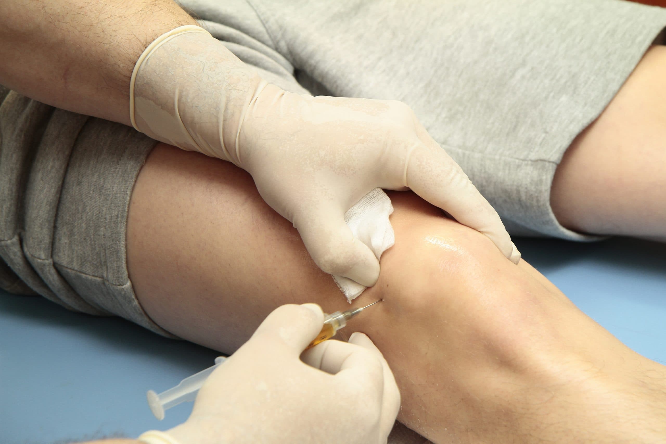


Close-up of knee assessment and treatment

Plica

From £9,800
View Price List

Guide price only. Final cost is confirmed after assessment.

A plica is a fold of tissue inside the joint that can become inflamed, thickened, or mechanically irritating. When a symptomatic plica causes persistent pain, clicking, or catching that has not responded to conservative treatment, arthroscopic excision offers a targeted, minimally invasive solution. At London Cartilage Clinic, plica surgery is performed as a day-case keyhole procedure with rapid recovery.

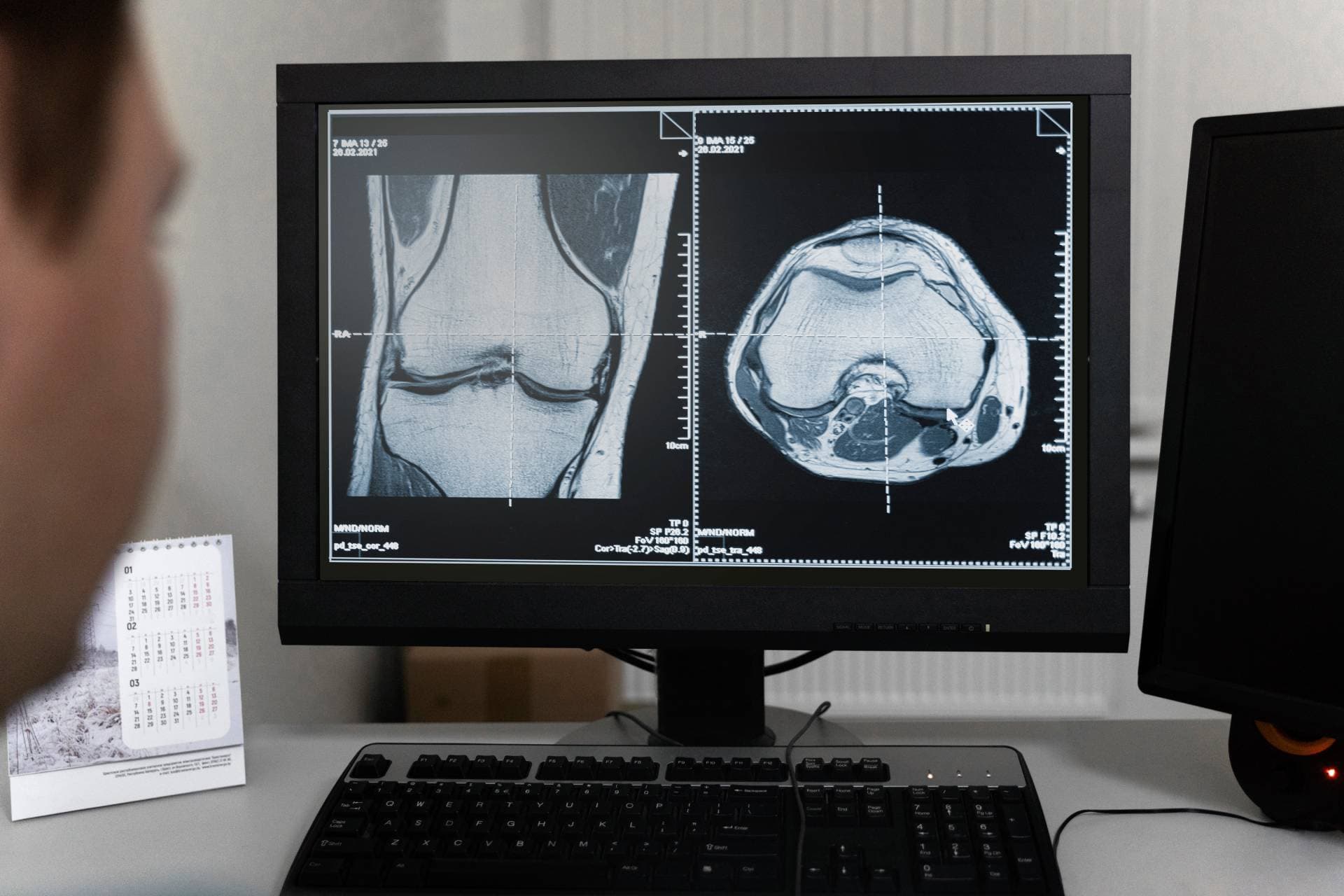
Knee MRI scans being reviewed on a monitor

When a plica becomes a problem

Plicae are a normal part of joint anatomy in most people. They only become clinically significant when they thicken, scar, or become caught between the moving surfaces of the joint during bending and straightening.

  • The medial plica of the knee is the most commonly symptomatic location.
  • Symptoms often mimic meniscal tears or patellofemoral pain, making diagnosis challenging.
  • A chronically irritated plica can damage the cartilage surface beneath it if left untreated.

Accurate diagnosis is the most important step. Many patients with plica syndrome have been told their pain is from other causes. Specialist assessment can identify the plica as the source and guide appropriate treatment.

Arthroscopic excision and rapid recovery

When conservative treatment has not resolved symptoms, arthroscopic plica excision removes the offending tissue through a minimally invasive approach.

  • Day-case procedure with small keyhole incisions.
  • Immediate weight-bearing after surgery in most cases.
  • Return to normal activity within two to four weeks.

At the time of arthroscopy, the surgeon also assesses the rest of the joint to check for any associated cartilage or meniscal damage that may need attention.

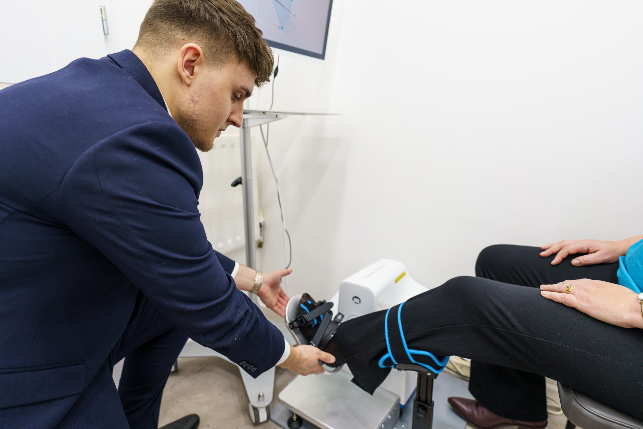
Knee rehabilitation after surgery

You may have more options than you think

Most patients have more treatment options than they have been told

At London Cartilage Clinic we follow a structured clinical framework across four areas of treatment. Before recommending a single procedure, we assess which combination of approaches gives you the best outcome.

Preserve

Protect what you have. Slow degeneration and manage symptoms.

Repair

Fix specific damage. Torn tissue, unstable joints, structural problems.

Regenerate

Rebuild lost tissue. Biological treatments that stimulate new growth.

Replace

When other options are exhausted. Joint replacement as a last resort.

Explore the full range of treatments available for your joint. Each hub page shows every option we offer, organised by clinical approach.

consulting-in-office-with-pen

Frequently Asked Questions

What is a plica?

A plica is a fold of synovial tissue inside the joint, a remnant from embryological development. Most people have plicae that cause no symptoms at all. In some cases, however, a plica can become thickened, inflamed, or trapped between the joint surfaces, producing pain, clicking, and catching.

How is plica syndrome diagnosed?

Diagnosis is based on clinical examination, symptom pattern, and ruling out other causes of anterior knee or elbow pain. MRI can sometimes show a thickened plica, but many are only confirmed during arthroscopy. A specialist who understands plica presentations is important for avoiding misdiagnosis.

Does plica syndrome always need surgery?

No. Many cases respond to physiotherapy, activity modification, and anti-inflammatory treatment. Surgery is considered when symptoms persist despite conservative management, particularly if the plica is causing mechanical catching or is damaging the underlying cartilage surface.

What does the surgery involve?

Arthroscopic plica excision is a keyhole procedure. The thickened or inflamed plica is identified and removed or debrided using small instruments inserted through two to three tiny incisions. The procedure is typically performed as a day case under general or regional anaesthetic.

What is the recovery time?

Recovery after plica excision is usually rapid. Most patients can weight-bear immediately and return to normal activities within two to four weeks. Physiotherapy may be recommended to address any muscle weakness or movement patterns that developed while the plica was symptomatic.

Still have more specific concerns?

Free Discovery Call

London Cartilage Clinic

Latest Insights

Clinical updates, cartilage treatment guidance, and recovery-focused articles from our specialist team.

When hip pain at night needs a specialist
Joint Conditions
Eleanor Hayes

When hip pain at night needs a specialist

Lying down removes the protective effects of movement and muscle engagement, concentrating pressure on already-affected joint surfaces for hours at a time. Where pain occurs — outer hip, deep groin, or radiating leg — identifies the underlying cause without imaging.

ChondroFiller plus Arthrosamid for advanced knee OA
ChondroFiller / Liquid Cartilage
Eleanor Hayes

ChondroFiller plus Arthrosamid for advanced knee OA

Advanced knee osteoarthritis typically has two concurrent pain sources—cartilage wear and synovial inflammation—that conventional single-agent injections address incompletely. The CFI+ protocol treats both by placing ChondroFiller, a collagen scaffold, on the damaged cartilage and injecting Arthrosamid, a polyacrylamide hydrogel, into the joint lining in one outpatient appointment.

ChondroFiller Injection for Thumb Base Cartilage Damage
Cartilage Repair
Eleanor Hayes

ChondroFiller Injection for Thumb Base Cartilage Damage

Standard steroid injections at the thumb base provide four to five months of relief, with effects diminishing on repeat use; ChondroFiller, an injectable collagen scaffold administered as an outpatient, recruits the body's own progenitor cells to repair cartilage, with prospective evidence of sustained improvements over two years and no adverse events.

Privacy & Cookies Policy