


ChondroFiller / Liquid Cartilage
Eleanor Hayes
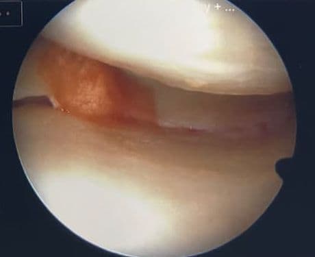
IKDC and MOCART scores after ChondroFiller injection
Functional recovery from ChondroFiller outpaces structural repair: patients achieve a clinically meaningful 30-point IKDC improvement within twelve months, but cartilage maturation on MRI continues beyond that functional plateau.

