
Cartilage Repair
Eleanor Hayes
Single-Session Cartilage Repair with STACi
Cartilage repair that once required two operations spanning three to six months is now completed in a single operative session lasting 2.5–4 hours through STACi.

Guide price only. Final cost is confirmed after assessment.
Sub-chondroplasty is a targeted procedure that treats bone marrow lesions (BMLs), areas of damaged bone beneath the cartilage surface that are a recognised source of deep, persistent joint pain. A flowable calcium phosphate bone substitute is injected directly into the lesion under fluoroscopic guidance, filling the defect and providing a scaffold for new bone to form. At London Cartilage Clinic, sub-chondroplasty is often combined with arthroscopy to address both the bone and cartilage components of your condition.
 Reviewed byProf Paul Lee MBBch, FRCS (Tr & Orth), PhDLast reviewed 1 May 2026
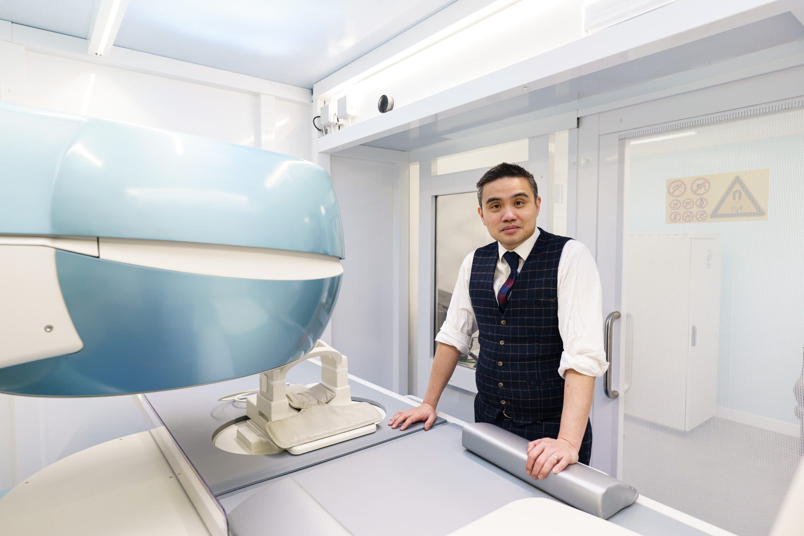
The subchondral bone sits directly beneath the cartilage and plays a critical role in distributing load across the joint. When this bone is damaged, it becomes a source of deep, aching pain that does not respond to surface-level treatments.
Identifying the bone marrow lesion as the pain source is key. Patients are often surprised that their pain is coming from the bone rather than the cartilage, and that a targeted treatment exists.
Sub-chondroplasty is performed under image guidance. The bone substitute is delivered through a cannula placed directly into the lesion, filling the defect from within.
Sub-chondroplasty can provide meaningful pain relief for patients whose symptoms are driven by subchondral bone pathology rather than cartilage damage alone.

You may have more options than you think
At London Cartilage Clinic we follow a structured clinical framework across four areas of treatment. Before recommending a single procedure, we assess which combination of approaches gives you the best outcome.
Protect what you have. Slow degeneration and manage symptoms.
Fix specific damage. Torn tissue, unstable joints, structural problems.
Rebuild lost tissue. Biological treatments that stimulate new growth.
When other options are exhausted. Joint replacement as a last resort.
Explore the full range of treatments available for your joint. Each hub page shows every option we offer, organised by clinical approach.

A bone marrow lesion (BML) is an area of abnormal signal seen on MRI in the subchondral bone, just beneath the cartilage surface. It represents fluid accumulation, micro-damage, or altered bone metabolism. BMLs are a recognised source of deep, persistent joint pain, particularly in osteoarthritis.
A flowable calcium phosphate bone substitute is injected directly into the bone marrow lesion under fluoroscopic guidance. The material fills the defect, provides structural support, and acts as a scaffold for new bone formation. The procedure can be performed alongside arthroscopy.
Sub-chondroplasty is suited to patients with MRI-confirmed bone marrow lesions that cause pain out of proportion to the cartilage damage visible on imaging. It is often considered when injections and physiotherapy have not adequately controlled symptoms, but the patient is not ready for or does not need joint replacement.
The procedure can be performed under general or regional anaesthetic. It is often combined with an arthroscopy to assess and treat any associated cartilage or meniscal pathology at the same time.
Most patients use crutches for two to four weeks with progressive weight-bearing. Pain from the bone marrow lesion typically improves over the following weeks as the bone substitute incorporates. Full recovery to normal activity is expected within two to three months.
Still have more specific concerns?
Free Discovery CallLondon Cartilage Clinic
Clinical updates, cartilage treatment guidance, and recovery-focused articles from our specialist team.
Cartilage repair that once required two operations spanning three to six months is now completed in a single operative session lasting 2.5–4 hours through STACi.

A partial ACL tear does not always require surgery; the decision hinges on whether the knee remains mechanically stable under load, though approximately 40% of young athletes managed conservatively eventually sustain a complete rupture.

ChondroFiller hip injections cost £6,500–£9,500 and recruit the patient's own cells to repair cartilage; a structured preservation model requires yearly imaging and bi-annual top-up injections.