

The knee is an engineering marvel, built to provide stability, flexibility, and strength —qualities we need for everything from everyday walking to high-level sports. Central to the knee ’s stability is the anterior cruciate ligament, or ACL, which helps keep the joint steady and prevents movements that could cause injury. Understanding how the knee is built—and how it functions—is essential to grasping how ACL injuries occur and how best to treat them. In this article, we’ll explore the knee’s intricate structure, how subtle anatomical differences can affect ACL injury risk, and why this knowledge is crucial for effective treatment.
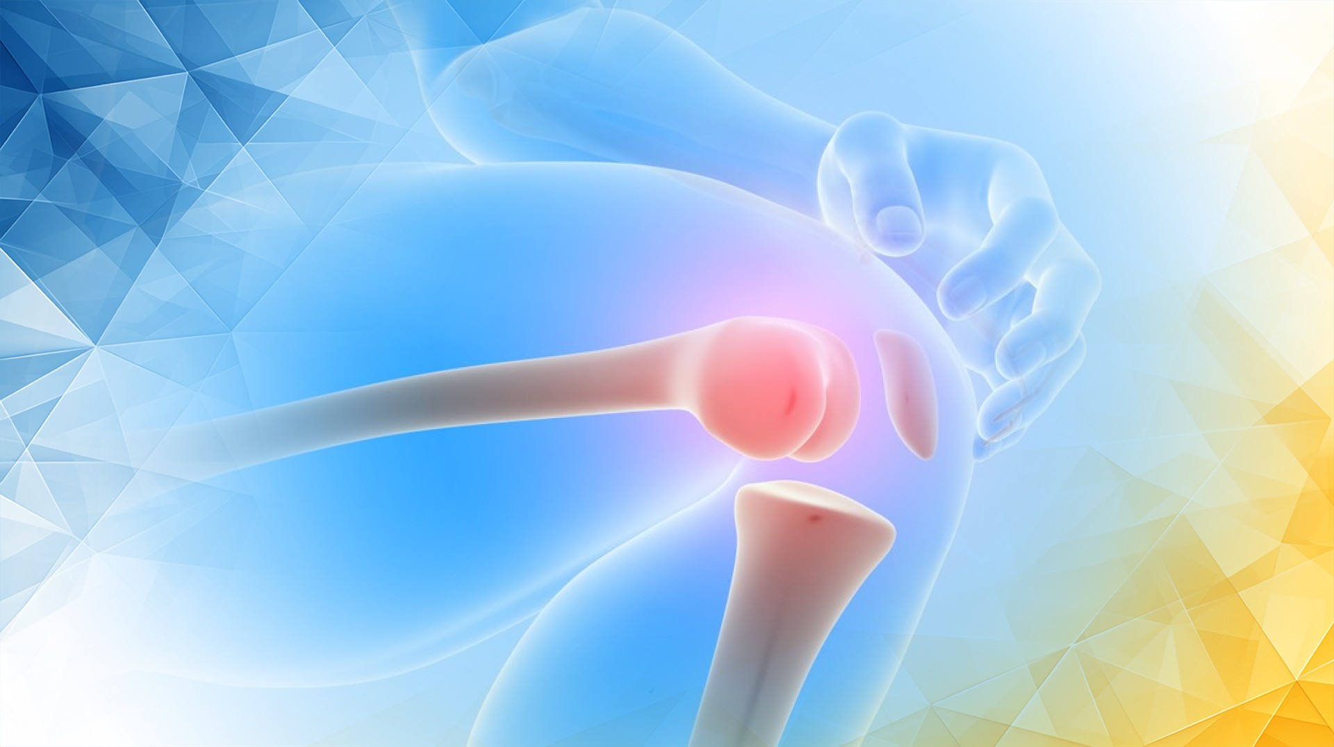
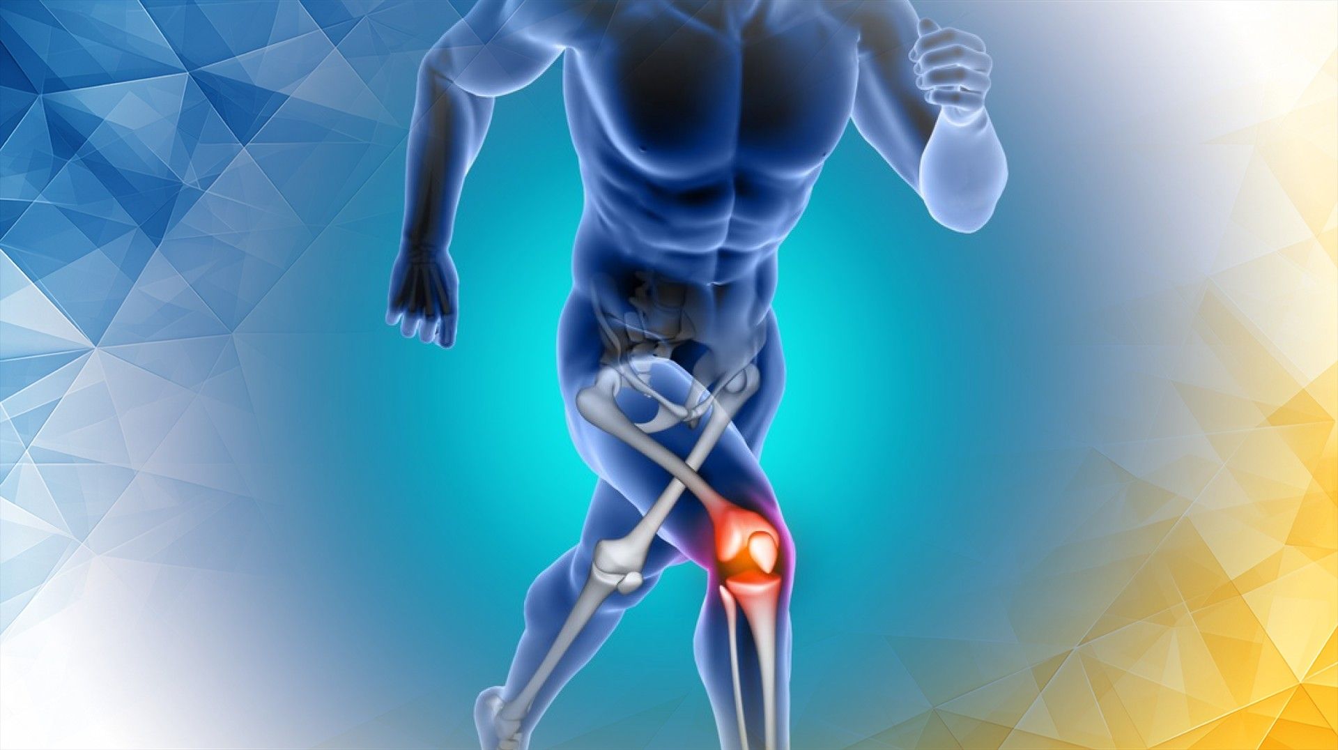
The knee joint connects the thigh bone (femur) to the shin bone ( tibia ) and is held together by an intricate network of ligaments, tendons, muscles, and cartilage. Four major ligaments act as stabilizers: the ACL, posterior cruciate ligament (PCL), medial collateral ligament (MCL), and lateral collateral ligament (LCL). The menisci—two C-shaped pieces of cartilage —serve as shock absorbers, distributing weight and cushioning the joint.
All these components work together seamlessly to keep the knee stable during movements like bending, twisting, or changing direction. Understanding this interplay is key to recognizing what goes wrong during an injury .
Knee anatomy isn’t just textbook knowledge. As Hassebrock et al. (2020) emphasize, having a solid understanding of knee ligament anatomy and biomechanics is foundational for physicians treating knee injuries, especially more complex or severe ones. The back of the knee, as LaPrade et al. (2007) explain, is especially complex, featuring a dense network of ligaments and soft tissues that are critical for stability.
The ACL is a tough, fibrous band that runs inside the knee, connecting the femur to the tibia. Its main job is to prevent the shin bone from sliding too far forward relative to the thigh bone and to control excessive twisting of the knee. This makes the ACL vital for activities that involve sudden stops, pivots, or jumps.
The ligament’s fibers extend from the lower end of the femur to the upper part of the tibia in a way that allows them to absorb and resist dangerous forces. However, under the right (or wrong) conditions, these forces can exceed what the ACL can withstand, leading to a tear.
Biomechanics play a big role here. As described by Hassebrock et al. (2020), understanding how the main knee ligaments function helps inform surgical and rehabilitative treatment choices. The complexity and importance of structures at the back of the knee, highlighted by LaPrade et al. (2007), further emphasize why a deep knowledge of knee anatomy is essential for advances in care.
No two knees are exactly alike—and even small differences in anatomy can have a major impact on ACL injury risk. For example, the slope of the tibial plateau (the top of the shin bone) can vary, and a steeper slope can increase forward pressure during activity, putting more strain on the ACL. Likewise, the intercondylar notch—a groove in the femur through which the ACL passes—can be wider or narrower from person to person. A narrow notch leaves less space for the ligament , increasing the risk of it being pinched or damaged.
Other differences, such as the precise angle of ACL fibers or variations in bone shape, can also influence susceptibility to tears. These anatomical quirks help explain why some people are more likely to suffer ACL injuries than others, even when doing similar activities.
Recognizing these variations is important for preventing injuries and for personalizing treatment. As Hassebrock et al. (2020) note, subtle differences in cruciate and collateral anatomy directly influence how the knee functions and responds to stress. Continuing research, as LaPrade et al. (2007) suggest, is helping us better understand the practical effects of these differences—improving both prevention and treatment strategies.
Knee anatomy isn’t just interesting—it directly shapes how ACL injuries are treated. When deciding between surgery and non-surgical approaches like physical therapy, doctors take into account both a patient’s lifestyle and the unique structure of their knee.
During ACL reconstruction surgery, surgeons aim to position the new graft as closely as possible to the original ligament, matching its placement and fiber direction. Doing this successfully is key to restoring normal knee movement and long-term stability. If the graft placement doesn’t account for individual anatomical differences, problems such as persistent instability or early joint wear can result.
A deeper understanding of the relationship between structure and function helps clinicians tailor treatments—there’s no one-size- fits -all solution for ACL injuries. For less active patients or those with certain knee shapes, a tailored rehab program may offer excellent results without surgery. When treatment matches both anatomy and personal needs, recovery is smoother, and the risk of reinjury drops dramatically.
Advances in imaging and surgical techniques are transforming how ACL injuries are diagnosed and repaired. High-definition MRI and 3D imaging let doctors study every detail of a patient’s knee, helping plan surgeries with remarkable precision.
New tools and computer-assisted methods enable surgeons to recreate the ACL’s natural position more accurately than ever before. Rehabilitation programs are also becoming more personalized, using detailed anatomical and biomechanical data to design recovery plans that fit a patient’s unique knee.
All these innovations are paving the way toward fully customized care, promising better outcomes and quicker, safer returns to activity.
In summary, understanding knee anatomy —especially the structure and function of the ACL—is central to identifying who is at risk for injury, how severe those injuries might be, and which treatments will work best. Even small differences in knee structure can have a big impact on both injury risk and recovery process.
As research and technology advance, personalized, anatomy-based care is becoming the new norm for ACL injury treatment. By appreciating the unique features of each knee, doctors can improve prevention, deliver more effective treatment, and help patients enjoy active, healthy lives for years to come.
Blackburn, T. A., & Craig, E. (1980). Knee anatomy. Physical Therapy, 60(12), 1556-1560. https://doi.org/10.1093/ptj/60.12.1556
Hassebrock, J. D., Gulbrandsen, M. T., Asprey, W. L., Makovicka, J. L., & Chhabra, A. (2020). Knee ligament anatomy and biomechanics. Sports Medicine and Arthroscopy Review, 28(3), 80-86.
LaPrade, R. F., Morgan, P. M., Wentorf, F. A., Johansen, S., & Engebretsen, L. (2007). The anatomy of the posterior aspect of the knee. The Journal of Bone and Joint Surgery (American), 89(4), 758-764. https://doi.org/10.2106/jbjs.f.00120
All our treatments are selected to help patients achieve the best possible outcomes and return to the quality of life they deserve. Get in touch if you have any questions.
At London Cartilage Clinic, we are constantly staying up-to-date on the latest treatment options for knee injuries and ongoing knee health issues. As a result, our patients have access to the best equipment, techniques, and expertise in the field, whether it’s for cartilage repair, regeneration, or replacement.
For the best in patient care and cartilage knowledge, contact London Cartilage Clinic today.
At London Cartilage Clinic, our team has spent years gaining an in-depth understanding of human biology and the skills necessary to provide a wide range of cartilage treatments. It’s our mission to administer comprehensive care through innovative solutions targeted at key areas, including cartilage injuries. During an initial consultation, one of our medical professionals will establish which path forward is best for you.
Contact us if you have any questions about the various treatment methods on offer.
Legal & Medical Disclaimer
This article is written by an independent contributor and reflects their own views and experience, not necessarily those of londoncartilage.com. It is provided for general information and education only and does not constitute medical advice, diagnosis, or treatment.
Always seek personalised advice from a qualified healthcare professional before making decisions about your health. londoncartilage.com accepts no responsibility for errors, omissions, third-party content, or any loss, damage, or injury arising from reliance on this material. If you believe this article contains inaccurate or infringing content, please contact us at [email protected].