sunset-bike-rest-country-road

Kneecap

The London Cartilage Clinic provides treatment for kneecap injuries and kneecap related injuries that are market leading. We have a range of services to cater to a range of patients and circumstances.

MPFL repair illustration

MPFL repair

Dislocation of the kneecap can be a result of damage to medial patellofemoral ligament (MPFL). The London Cartilage Clinic has the resources and expertise to repair this ligament and prevent future kneecap dislocations.

Learn More
Chondromalacia illustration

Chondromalacia

A Chondromalacia affliction can result in the weakening of the cartilage behind the kneecap. This leads to an increased likelihood of tearing and dislocation. Fortunately, we have dedicated treatments to fix this problem.

Learn More
Osteotomy illustration

Osteotomy

Surgery can be undertaken to remove small parts of the bone (or bones) in the knee joint, thereby allowing the patella to fit comfortably in its proper position. A knee osteotomy is considered a major procedure, so it’s important patients are fully aware of what is involved and the potential complications.

Learn More
Joint replacement illustration

Joint Replacement

Our specialist surgeons can complete a knee arthroplasty, where part of the kneecap and other bones in the joint may be replaced by artificial material. This is an effective treatment option for dislocated knees and osteoarthritis.

Learn More
Professor in brown plaid suit discussing kneecap anatomy

What is the kneecap?

The patella (kneecap) is a small, roughly triangular shaped bone that makes up part of the knee joint. It serves to protect the soft tissues and leg bones that are responsible for allowing the knee to bend. However, the kneecap also provides additional leverage for the leg muscles responsible for dictating knee motion. The back side of the patella is covered by a layer of cartilage which allows it to slide across the thighbone as the knee flexes.

Symptoms of kneecap dislocation

A dislocated kneecap is where the patella is moved out of its usual place where the femur, tibia, and fibula meet. It is usually caused by an impact, such as from a fall or a vehicle collision. If you have a dislocated knee, you are likely to experience the following symptoms:

  • Visual deformity.
  • The kneecap is moved to the side of the knee.
  • You are unable to straighten the knee.
  • Swelling, tenderness, and pain.
  • Inability to walk.
Doctor reviewing kneecap scan on a monitor
Modern operating room for kneecap dislocation treatment

Treatment options for kneecap dislocation

There are many options available when it comes to kneecap dislocation treatment which vary in their application. These range from kneecap dislocation surgery to targeted repair procedures and treatments to strengthen the tissue surrounding the patella. Regardless, a healthcare professional will first assess the severity of the dislocation to advise accordingly. The types of knee dislocation are medial, lateral, rotary, anterior, and posterior.

How can London Cartilage Clinic help?

By adhering to the highest standards of surgical practices, we ensure our patients' kneecap dislocation recovery time is as short as possible. We are constantly seeking the best ways to treat cartilage damage throughout the body, but especially to key areas such as the kneecap. As such, we have refined our treatments and surgical approach over time to ensure we always deliver the best results and experience for our patients.

Specialist kneecap treatment planning and surgical expertise
consulting-in-office-with-pen

Frequently Asked Questions

What common kneecap issues are treated at the clinic?

We treat a variety of kneecap issues, including patellofemoral pain syndrome, chondromalacia patellae, dislocations, and fractures. Our treatments range from physiotherapy and bracing, key-hole STARRs internal bracing to complex surgical interventions, depending on the severity and nature of the condition.

How does the clinic diagnose kneecap problems?

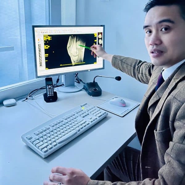
At the London Cartilage Clinic, the diagnosis of kneecap problems begins with a thorough clinical assessment, which includes a detailed review of the patient’s medical history and a comprehensive physical examination. To gain an accurate understanding of the condition, we employ advanced imaging technologies such as MRI and CT scans, which provide detailed insights into the state of the kneecap and the surrounding structures. A traditional X-ray can only provide a very limited information. For more complex cases, where standard imaging might not suffice, our clinic offers an innovative in-clinic procedure known as dynamic needle arthroscopy. This minimally invasive technique allows us to assess the internal structure of the knee joint in real time, providing us with a more precise and direct evaluation. This comprehensive approach ensures accurate diagnosis, which is critical for developing effective treatment plans tailored to each patient’s specific needs.

What are the treatment options for kneecap pain or instability?

Treatment options vary based on the individual’s condition but can include physiotherapy, orthotics, pain management, and in some cases, surgery. Our focus is always on minimally invasive techniques and regenerative treatments to preserve as much natural structure and function as possible.

Still have more specific concerns?

Free Discovery Call

London Cartilage Clinic

Latest Insights

Clinical updates, cartilage treatment guidance, and recovery-focused articles from our specialist team.

Nutrition Strategies for Strengthening Knee Cartilage
John Davies

Nutrition Strategies for Strengthening Knee Cartilage

Discover how nutrition supports knee cartilage health, as explained by Professor Paul Lee of the London Cartilage Clinic. Learn which foods—like fatty fish, turmeric, and vitamin C-rich fruits—can strengthen cartilage and reduce inflammation. Avoid processed sugars and trans fats that harm joints. Practical meal tips help incorporate these nutrients into daily life. While diet aids joint comfort and resilience, it works best combined with exercise and medical advice. Embrace balanced eating and expert guidance for healthier, more comfortable knees.

ChondroFiller Minimally Invasive Cartilage Repair Benefits and Patient Suitability
John Davies

ChondroFiller Minimally Invasive Cartilage Repair Benefits and Patient Suitability

ChondroFiller is a minimally invasive, keyhole surgery technique that repairs cartilage damage by injecting a collagen gel directly into the affected area. Suitable for patients with small to medium cartilage lesions, it promotes natural cartilage regeneration with less pain and faster recovery compared to traditional surgeries. The procedure involves arthroscopic guidance for precise gel placement, minimizing tissue damage and complications. Clinical studies report high patient satisfaction, improved joint function, and lasting benefits. Recovery typically includes tailored rehabilitation with reduced pain and a quicker return to activity. ChondroFiller offers a safe, effective alternative for cartilage repair, emphasizing simplicity and rapid healing. Consultation with healthcare professionals is essential to determine candidacy and ensure optimal outcomes.

The Link Between Segond Fracture and ACL Tears in Diagnosis and Treatment
John Davies

The Link Between Segond Fracture and ACL Tears in Diagnosis and Treatment

Explore the critical connection between Segond fractures and anterior cruciate ligament (ACL) tears, common yet often intertwined knee injuries especially among athletes. This article highlights how the Segond fracture—a small tibial bone break—serves as a key indicator of ACL damage. Advances in MRI and CT imaging aid in early and accurate diagnosis, leading to more comprehensive treatment strategies that address both bone and ligament injuries. Understanding this relationship improves patient outcomes, enhancing knee stability and function. Future research aims to refine detection and develop minimally invasive surgical techniques, helping patients recover faster and return to active lifestyles confidently.