

Beyond the Knee: Exploring the Calf Connection in Behind Knee Pain
Introduction
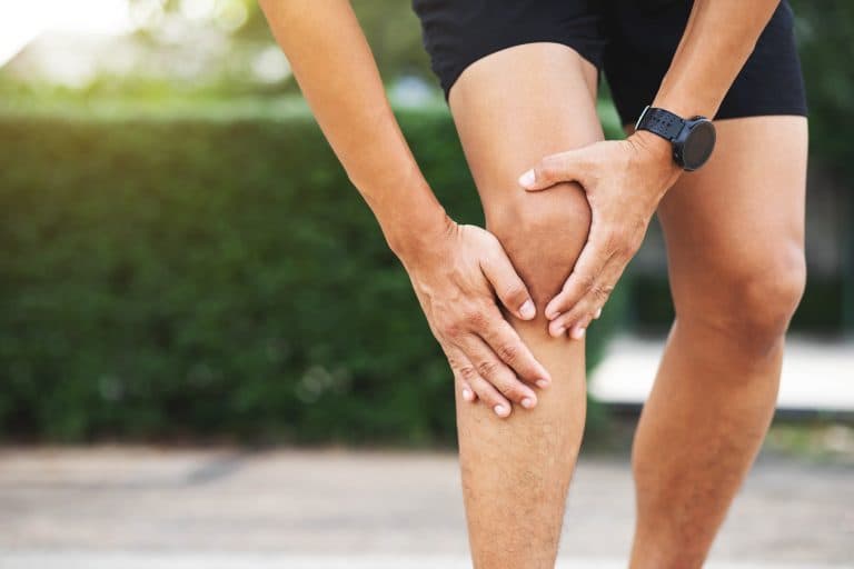
Pain behind the knee is something many people experience, regardless of age or activity level. While it’s easy to assume that all knee pain comes directly from the joint itself, discomfort in this area can significantly restrict movement and impact your daily life. In this article, we’ll explore how the calf muscles—the powerful muscles at the back of your lower leg—can directly influence pain behind the knee. Understanding this often-overlooked connection can help uncover the real source of your discomfort and lead to better treatment options.
Understanding the Calf-Knee Connection
To see why calf muscles matter when it comes to knee pain , it helps to know how closely connected these areas are. The muscles that support and move the knee include the calf muscles, specifically the gastrocnemius and soleus. These muscles originate just below the knee and play a big role in activities such as walking, running, and jumping. When calf muscles become tight, weak, or imbalanced, they can disrupt knee movement and place extra strain on the area behind the knee. This is often why pain in the back of the knee goes hand-in-hand with tightness or soreness in the upper calf.
How Calf Issues Can Cause Knee Pain
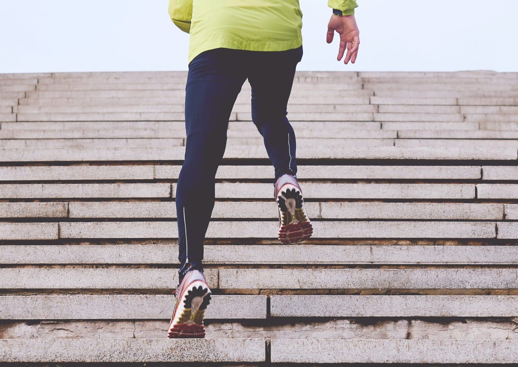
Problems in the calf muscles can lead to pain behind the knee in several ways. If the calf muscles are tired, weak, or inflexible, your body may compensate by moving in ways that put more pressure on the knee joint . High-impact activities like running or cycling can make this imbalance worse, since they require the calves to work hard. As a result, many people notice their knee pain increases after exercise or long periods of standing. During a check-up, a healthcare provider may find tightness in the calf, tenderness behind the knee, or reduced ankle mobility—all signs that the calves may be contributing to knee pain.
Next steps
Where to go from here
These routes are selected from the topic and purpose of this article. They are guidance, not a diagnosis or treatment recommendation.
Learn more
Explore treatment options
Talk to the team
Book a free discovery call
What the Research Tells Us
Research makes it clear that knee pain often involves more than the joint itself. Studies have found that discomfort can originate in areas surrounding the knee , including the soft tissues like the calf muscles. Addressing the health of these supporting muscles is an important piece of managing chronic or recurring knee issues. Evidence increasingly supports the idea that evaluating and treating both the knee and the nearby muscles—like the calves—leads to better outcomes. Healthcare professionals now routinely assess calf strength and flexibility when diagnosing knee pain, which has improved both diagnosis and treatment for patients.
How to Manage Pain Behind the Knee
The most effective approach to pain behind the knee focuses on both the knee joint and the calf muscles. Physiotherapy routines that include strengthening and stretching the calves can restore muscle balance and improve knee stability. Exercises such as calf raises, controlled lowering (eccentric movements), and calf stretches are particularly helpful. Supportive measures like knee braces or sleeves can also take pressure off the joint during activity. In addition to exercise, simple changes such as avoiding excessive overuse, warming up thoroughly, and gradually increasing activity levels can make a big difference. When patients and clinicians work together to target both calf strength and knee health, the chances of reducing pain and regaining comfortable movement are much greater.
Conclusion
The calf muscles are key players in many cases of pain behind the knee. Both clinical practice and scientific research highlight the strong connection between calf health and knee function . By taking a whole-body approach—one that recognises the importance of supporting muscles as well as the joint—it’s possible to achieve better, longer-lasting relief from knee pain . Understanding and addressing this calf-knee connection can lead to more effective treatment, improved mobility, and a better quality of life.
References
- Lewis, H. R., Chaplin, W. J., McWilliams, D. F., Millar, B. S., Shahtaheri, S. Y., Gladman, J. F. R., & Walsh, D. (2022). The association of painful and non-painful comorbidities with central mechanisms of knee pain. Annals of the Rheumatic Diseases, 81, 1822. https://doi.org/10.1136/annrheumdis-2022-eular.1586
- Sinclair, J., & Selfe, J. (2015). Sex differences in knee loading in recreational runners. Journal of Biomechanics, 48(10), 2171–2175. https://doi.org/10.1016/j.jbiomech.2015.05.016
- Smith, T. O., Drew, B., Meek, T. H., & Clark, A. (2013). Knee orthoses for treating patellofemoral pain syndrome. John Wiley & Sons, Ltd. https://doi.org/10.1002/14651858.cd010513
Next steps
Where to go from here
These routes are selected from the topic and purpose of this article. They are guidance, not a diagnosis or treatment recommendation.
Learn more
Explore treatment options
Use a structured overview to narrow down the most useful next step for your situation.
Talk to the team
Book a free discovery call
A non-medical call with the team to understand services and choose the right booking route.
Legal & Medical Disclaimer
This article is published by London Cartilage Clinic for general information and education only. It does not constitute medical advice, diagnosis, or treatment.
Always seek personalised advice from a qualified healthcare professional before making decisions about your health. London Cartilage Clinic accepts no responsibility for errors, omissions, third-party content, or any loss, damage, or injury arising from reliance on this material.
If you believe this article contains inaccurate or infringing content, please contact us at [email protected].


