
Cartilage Repair
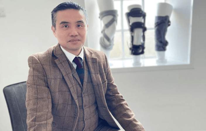
Eleanor Hayes
Single-Session Cartilage Repair with STACi
Cartilage repair that once required two operations spanning three to six months is now completed in a single operative session lasting 2.5–4 hours through STACi.

Femoroacetabular impingement occurs when abnormal bone shape at the hip creates friction during movement, damaging the labrum and cartilage. Early recognition and treatment can prevent progressive joint damage.
 Reviewed byProf Paul Lee MBBch, FRCS (Tr & Orth), PhDLast reviewed 1 May 2026
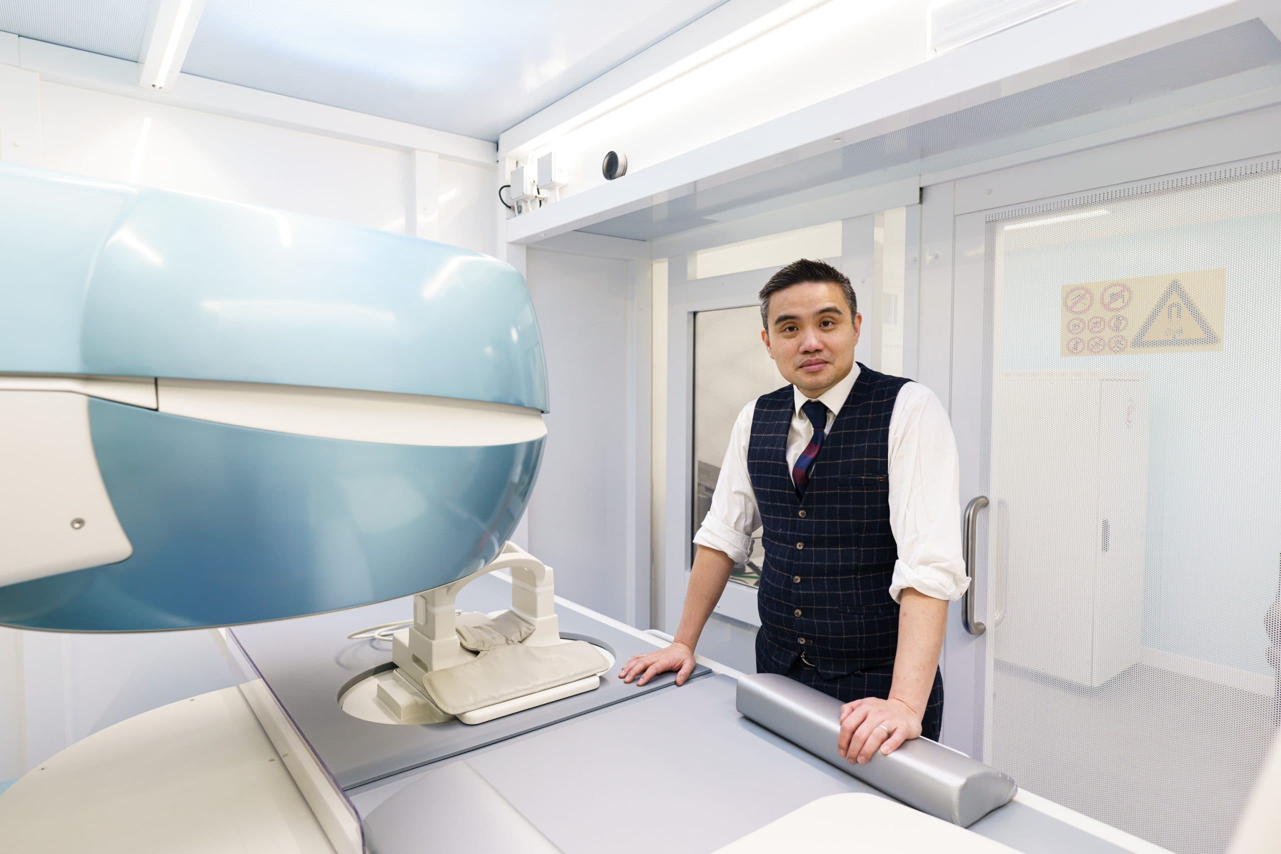
FAI is caused by abnormal bone morphology that develops during growth. The bone shape creates mechanical conflict between the femur and the socket during everyday hip movements, particularly flexion and rotation.
FAI typically presents with:
Diagnosis is confirmed with plain X-rays to assess bone morphology and MRI arthrogram to evaluate the labrum and cartilage.

Treatment is tailored to the severity of impingement and the degree of soft tissue damage. Our hip specialists offer both conservative and surgical pathways.
For patients with progressive labral damage or cartilage wear, hip arthroscopy to reshape the bone and repair the labrum addresses both the cause and the consequence of impingement. The goal is to restore pain-free hip movement and protect the joint from further degeneration.
You may have more options than you think
At London Cartilage Clinic we follow a structured clinical framework across four areas of treatment. Before recommending a single procedure, we assess which combination of approaches gives you the best outcome.
Protect what you have. Slow degeneration and manage symptoms.
Fix specific damage. Torn tissue, unstable joints, structural problems.
Rebuild lost tissue. Biological treatments that stimulate new growth.
When other options are exhausted. Joint replacement as a last resort.
Explore the full range of treatments available for your joint. Each hub page shows every option we offer, organised by clinical approach.

FAI is a condition where abnormal bone shape at the hip joint causes the femur (thigh bone) and acetabulum (socket) to make contact during normal movement. This repeated friction damages the labrum and cartilage over time, leading to pain and potentially early arthritis.
Cam impingement involves a bump on the femoral head that jams into the socket during flexion. Pincer impingement involves excess bone on the socket rim that grips the femoral neck. Many patients have a combination of both, known as mixed impingement.
Not always. Mild FAI with manageable symptoms may respond to physiotherapy and activity modification. However, if the impingement is causing progressive labral or cartilage damage, arthroscopic surgery to reshape the bone and repair the labrum may be recommended to protect the joint long-term.
Hip arthroscopy is used to reshape the abnormal bone (osteoplasty) and repair the damaged labrum. The procedure addresses both the cause (bone shape) and the consequence (soft tissue damage) in a single operation.
Still have more specific concerns?
Free Discovery CallLondon Cartilage Clinic
Clinical updates, cartilage treatment guidance, and recovery-focused articles from our specialist team.
Cartilage repair that once required two operations spanning three to six months is now completed in a single operative session lasting 2.5–4 hours through STACi.

A partial ACL tear does not always require surgery; the decision hinges on whether the knee remains mechanically stable under load, though approximately 40% of young athletes managed conservatively eventually sustain a complete rupture.

ChondroFiller hip injections cost £6,500–£9,500 and recruit the patient's own cells to repair cartilage; a structured preservation model requires yearly imaging and bi-annual top-up injections.