Introduction
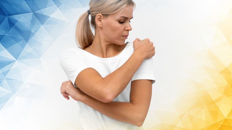
Knee ligament injuries are common, especially among athletes and active individuals. Of these, anterior cruciate ligament (ACL) tears are particularly significant because they often cause pain, instability, and a noticeable impact on movement and sports performance. Treating ACL injuries requires careful attention and rehabilitation, making them a major focus in sports medicine. While the knee contains several stabilizing ligaments—such as the medial collateral ligament (MCL)—this article will focus on the ACL: how tears occur, how they are diagnosed, and the latest treatments helping patients get back on their feet.
Knee Anatomy and Ligament Function
The knee is a complex joint stabilized by several strong ligaments. Two of the most important are the ACL and the MCL. The ACL runs diagonally inside the knee, connecting the thigh bone (femur) to the shin bone (tibia), and acts as the knee’s main stabilizer for forward movement and rotation. The MCL, found along the inner side of the knee, prevents the joint from buckling inward.
Think of the ACL as the knee’s “rotational anchor,” while the MCL functions as a “side brace.” Because the ACL controls complex twisting and forward movements, injuries to this ligament often cause more instability and tend to be more serious than MCL injuries.
How ACL Injuries Occur
ACL tears most often happen during activities that involve sudden stops, rapid direction changes, or awkward landings—frequent movements in sports like soccer, basketball, or skiing. For instance, if you cut sharply to the side or land off-balance after a jump, the shin bone may slide or twist too far forward, putting stress on the ACL until it tears.
Conversely, MCL injuries are usually caused by a direct blow to the outer knee, forcing it inward. Both ligaments can be injured at the same time, but ACL tears are especially common in sports that demand quick pivots and high-speed changes in movement.
Recent research has found that some valgus (inward-angling) injuries may tear both the MCL and a portion of the ACL, and that inflammation from a torn MCL may actually support healing of the ACL (Razi et al., 2020). Additionally, managing combined ACL- MCL injuries can be especially challenging because they may have a more complex prognosis than isolated ligament tears (Papalia et al., 2009).
Free non-medical discussion
Not sure what to do next?
Information only · No medical advice or diagnosis.
Diagnosing ACL Tears
Accurate diagnosis is key to proper treatment. Today, doctors rely mostly on imaging to determine the extent of an ACL injury . Magnetic resonance imaging (MRI) is the gold standard because it provides detailed pictures of the knee’s soft tissues, showing whether the ACL is partially or fully torn.
Other techniques, such as CT scans and arthroscopy (using a tiny camera to look inside the joint), can also help clarify the diagnosis. Advancements like 3D MRI give even clearer views, allowing doctors to assess the injury in greater detail and spot any related damage.
Experts agree, however, that more research is needed to determine the best diagnostic and treatment approaches, especially for combined ACL- MCL injuries (Papalia et al., 2009).

Treatment Options for ACL Tears
Treatment depends on the severity of the tear and the patient’s activity level. For small or partial tears, or for people who are less physically active, physical therapy and knee braces may restore stability without surgery.
A complete tear, especially in active individuals, often requires surgical reconstruction. Using minimally invasive arthroscopic techniques, surgeons reconstruct the torn ACL with a tissue graft taken from the patient or a donor. These advanced procedures mean less surgical trauma, less pain, and quicker recoveries than traditional open surgeries.
Interestingly, some studies have documented spontaneous healing in certain patients with both ACL and MCL tears , even without surgery (Razi et al., 2020). Research has also shown that when the MCL is treated nonoperatively—often with a brace or cast—before ACL reconstruction , patients do just as well as those with an isolated ACL tear , and there is no increased risk of ACL re- tearing . This means that unnecessary surgery and potential complications can sometimes be avoided (Bauman et al., 2024). However, there is still no universal agreement on the ideal treatment for combined injuries, and more research is needed (Papalia et al., 2009).
Rehabilitation and Recovery
Healing doesn’t stop after surgery or bracing—rehabilitation is essential to restore knee function . Physical therapy focuses on gradually improving range of motion, rebuilding strength , and retraining balance and coordination.
Rehabilitation programs are becoming more individualized, often using technology like biofeedback and even virtual reality to keep patients motivated and track progress. This personalized approach helps speed up recovery and ensures a safer return to daily activities or sports.
Conclusion and Looking Ahead
ACL tears present a real challenge due to their impact on knee stability and activity levels. Thanks to a deeper understanding of knee anatomy , injury mechanisms, advanced diagnostic imaging, and continual improvements in treatment and rehabilitation, managing ACL injuries has become more effective than ever.
New research and technologies on the horizon promise even better care for people with ACL injuries , offering hope for faster, stronger recoveries and less time spent sidelined from the activities they love.
References
Razi, M., Soufali, A. P., Ziabari, E. Z., Dadgostar, H., Askari, A., & Arasteh, P. (2020). Treatment of concomitant ACL and MCL injuries: Spontaneous healing of complete ACL and MCL tears. The Journal of Knee Surgery, 34(12), 1329–1336. https://doi.org/10.1055/s-0040-1708858
Bauman, S., Benner, R. W., & Shelbourne, K. D. (2024). Poster 341: Nonoperative treatment of MCL tears in ACL/MCL injuries does not increase ACL retear rates. Orthopaedic Journal of Sports Medicine, 12(7_suppl2). https://doi.org/10.1177/2325967124s00307
Papalia, R., Osti, L., Del Buono, A., Denaro, V., & Maffulli, N. (2009). Management of combined ACL-MCL tears: a systematic review. British Medical Bulletin, 93(1), 201-215. https://doi.org/10.1093/bmb/ldp044
Legal & Medical Disclaimer
This article is written by an independent contributor and reflects their own views and experience, not necessarily those of London Cartilage Clinic. It is provided for general information and education only and does not constitute medical advice, diagnosis, or treatment.
Always seek personalised advice from a qualified healthcare professional before making decisions about your health. London Cartilage Clinic accepts no responsibility for errors, omissions, third-party content, or any loss, damage, or injury arising from reliance on this material.
If you believe this article contains inaccurate or infringing content, please contact us at [email protected].
London Cartilage Clinic
Modern treatments, designed for lasting knee outcomes
Every treatment plan is selected to help you return to the quality of life you deserve. Our team combines up-to-date techniques with consultant-led decision making across cartilage repair, regeneration and replacement.
What your journey can look like
Step 01
Consultant-Led Assessment
Symptoms, imaging and goals reviewed in one structured appointment.
Step 02
Personalised Treatment Plan
A clear route chosen from repair, regeneration or replacement options.
Step 03
Precision Delivery
Modern techniques and specialist equipment matched to your diagnosis.
Step 04
Long-Term Knee Health
Follow-up guidance focused on durable function and quality of life.
Will the Arthrosamid work for me
PAAG-8+ Questionnaire
A bright, structured entry point for patients exploring Arthrosamid. The questionnaire helps frame suitability, expectations, and 24-month clinical benefit discussion before a fuller consultant-led review.
Designed for shared decision-making. It does not replace specialist consultation, examination, imaging review, or formal medical advice.