 Cartilage
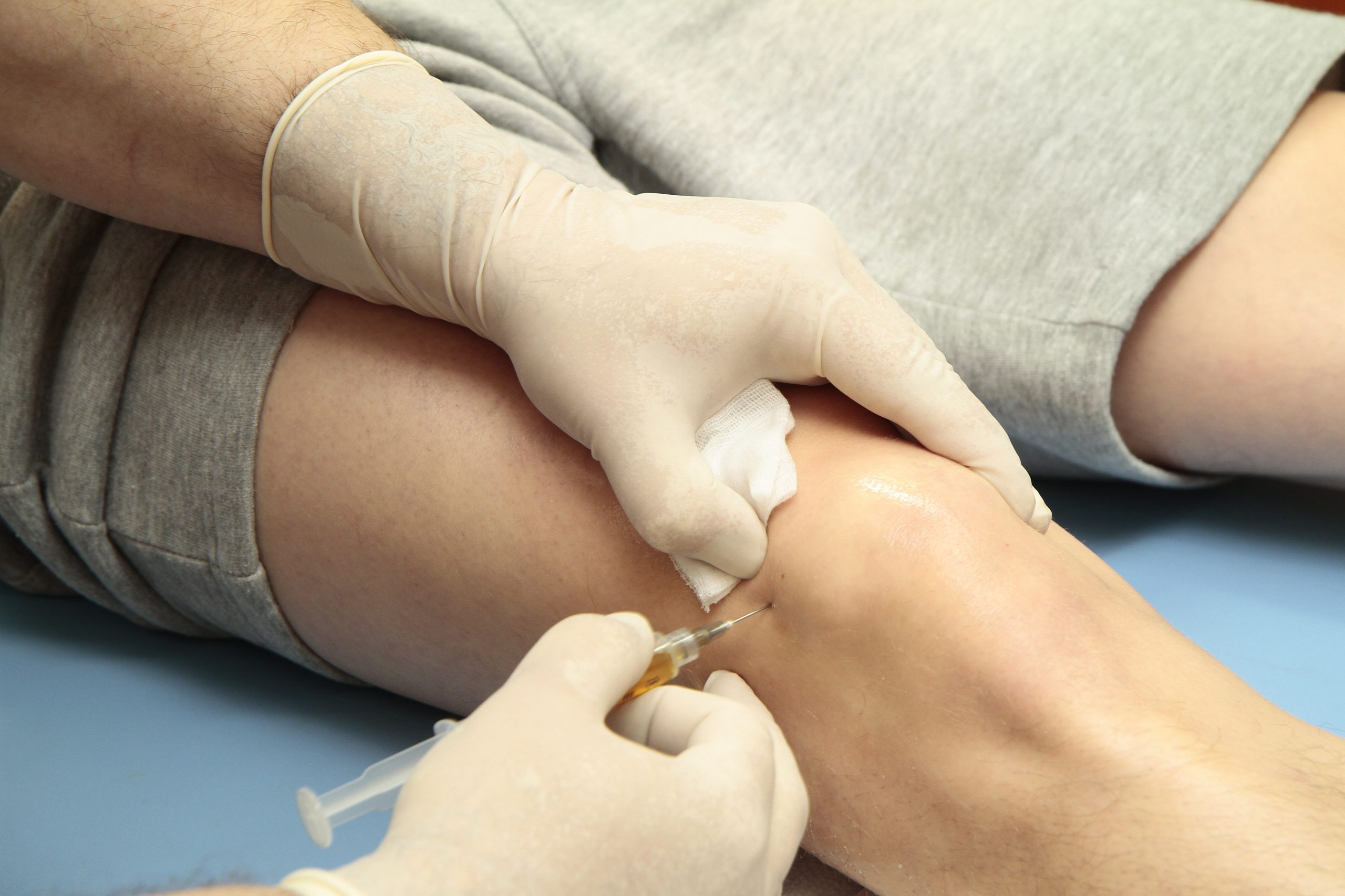 Knees
Knees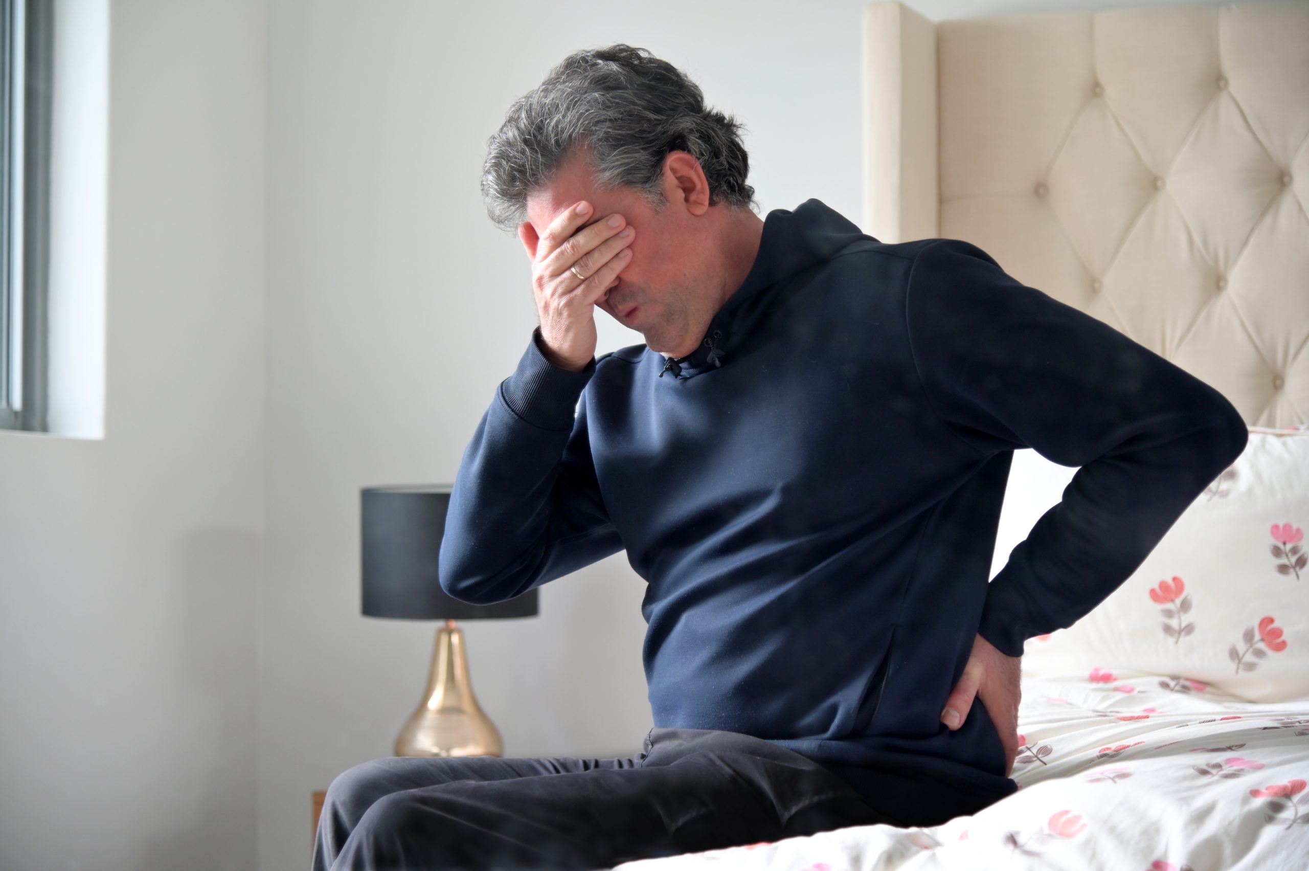 Hip
Hip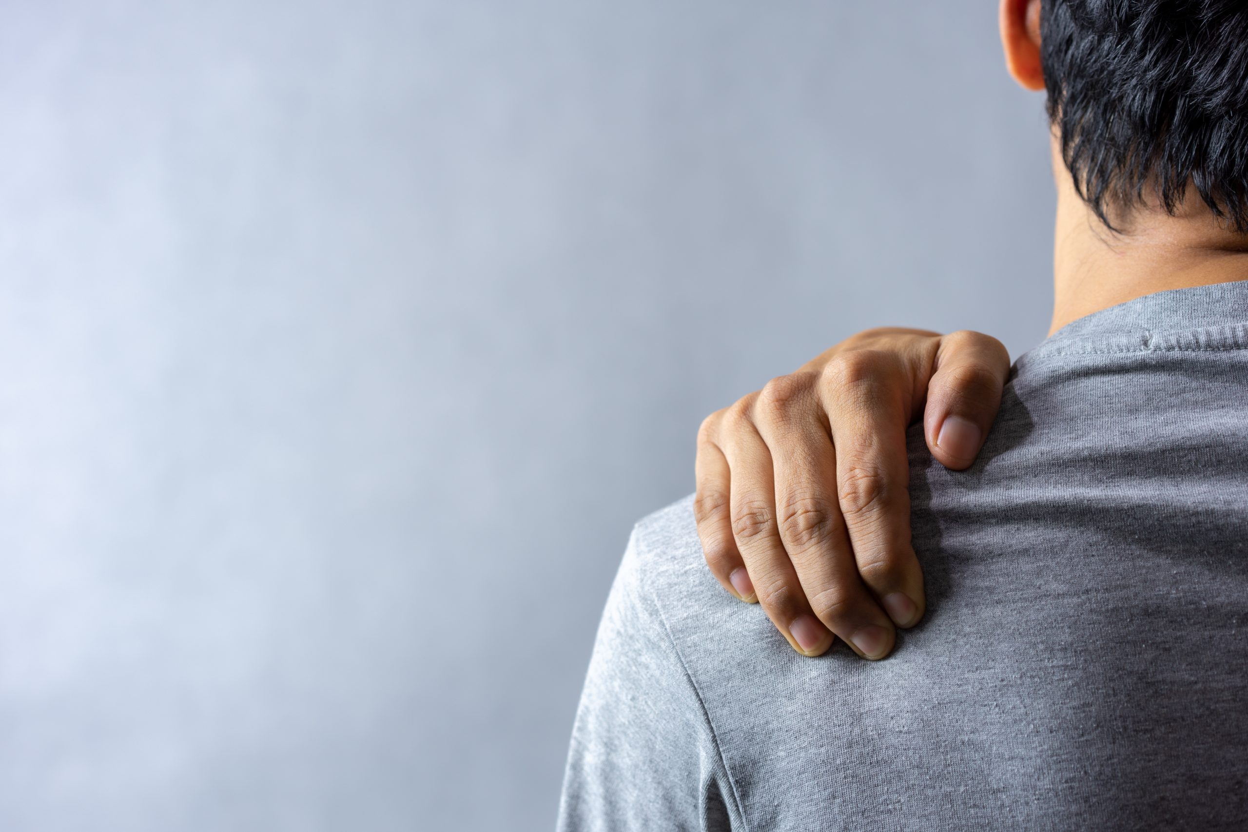 Shoulder
Knee Cartilage and Its Capacity for Repair
23/02/26


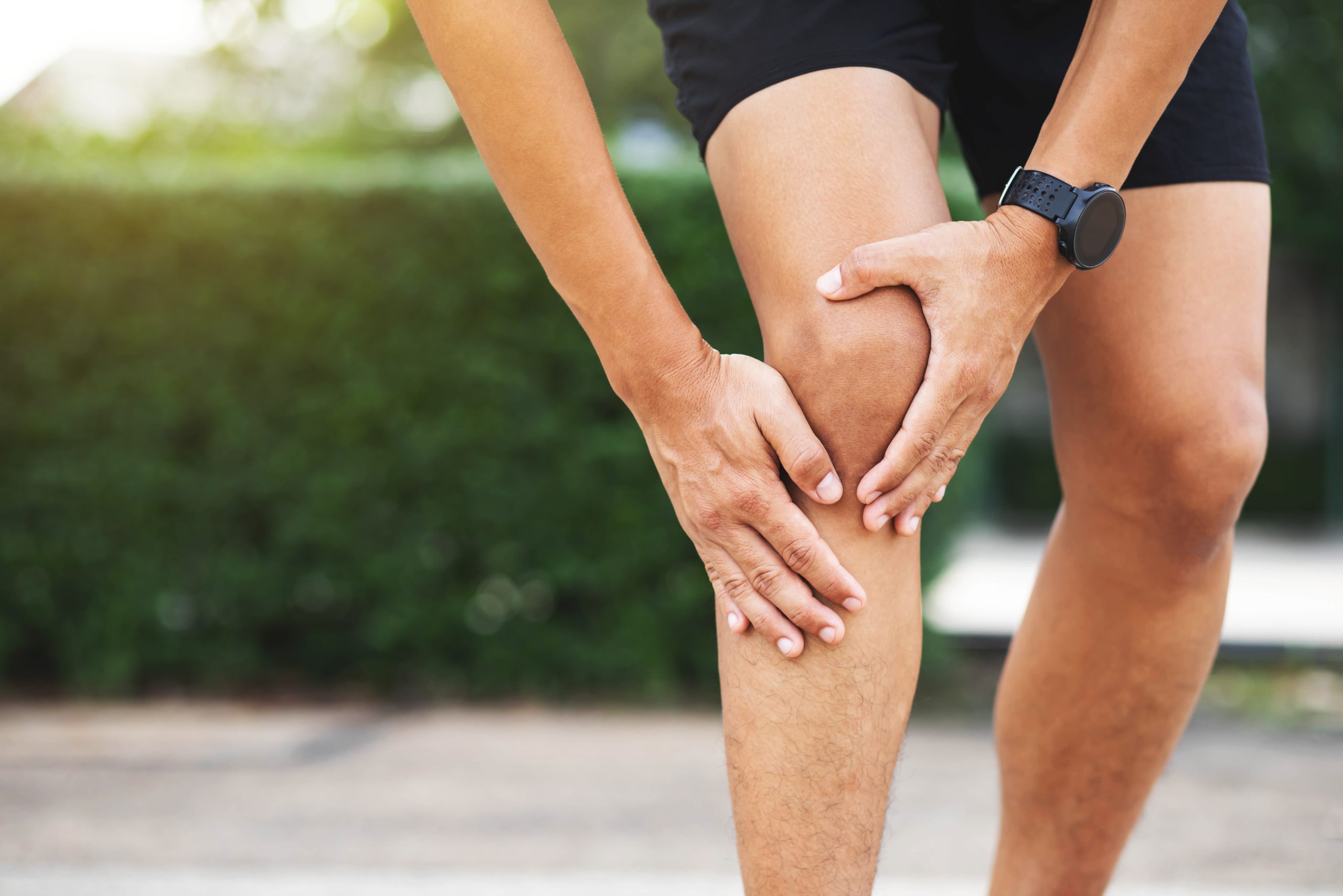
Articular cartilage is essential for smooth and pain-free movement within synovial joints, such as the knee. This specialised type of connective tissue covers the ends of bones in joints, allowing them to glide comfortably against one another while absorbing mechanical loads. Understanding what articular cartilage is, its role and how it functions is vital—especially since damage to this tissue can greatly affect joint health and mobility. In this article, we’ll explore the structure and function of articular cartilage, how injuries occur and cause pain, typical recovery times, and the latest treatment options on the horizon.
Articular cartilage is a specialised form of hyaline cartilage found specifically at joint surfaces. It is surprisingly thin—usually between 1 and 6 millimetres thick—yet its design is finely tuned for durability and flexibility. As Walter Herzog explains, “Articular cartilage is a thin (about 1–6 mm in human joints) layer of fibrous connective tissue covering the articular surfaces of bones in synovial joints” (Herzog, 2006). This tissue is made up mostly of cells called chondrocytes, which are embedded in a gel-like matrix rich in proteins such as collagen and proteoglycans. Together, these components help cartilage withstand the stresses of daily movement.
There are other types of cartilage too. Fibrocartilage, found in places like the discs between vertebrae and the menisci in the knee, is tougher and designed for shock absorption. Elastic cartilage, which contains elastic fibres, offers flexibility and is found in areas like the ears and the epiglottis. Interestingly, studies show that “thin split-thickness transplants of the cells of the gliding surface of immature articular cartilage induced the formation of fibrous tissue,” while thicker slices that include more active, ‘germinal’ cells can even stimulate new bone growth (Urist & Adams, 1968). This highlights how biologically active articular cartilage can be under certain conditions.
Injuries to articular cartilage can arise from sudden trauma, repetitive strain, or degenerative diseases such as osteoarthritis. When damaged, this tissue often causes symptoms like joint pain, swelling, stiffness, and decreased mobility. However, because the cartilage itself lacks nerves, the pain experienced usually comes from changes in other joint parts and increased friction between bones. As Herzog points out, “Osteoarthritis is a joint disease that is associated with a degradation and loss of articular cartilage from the joint surfaces and a concomitant increase in joint friction causing pain and disability, particularly in the elderly population” (Herzog, 2006). These symptoms often develop gradually, so early diagnosis can be challenging. Moreover, because cartilage receives very little blood flow, it has limited ability to heal on its own.
Advanced imaging techniques play a crucial role in assessing such injuries. Paunipagar and Rasalkar observe that “MRI has played a vital role in evaluation of articular cartilage” by revealing detailed images of both the microscopic and larger-scale structure of cartilage and helping detect early damage (Paunipagar & Rasalkar, 2014).
Recovery following cartilage injury varies widely depending on how severe the damage is, the patient’s lifestyle, and treatment choices. Typically, healing can take anywhere from several weeks to months. Many patients begin with conservative, non-surgical care.
These non-invasive approaches often involve physiotherapy to strengthen muscles around the joint, modifications in activity to ease joint stress, and medication to reduce pain and inflammation. The goal is to support healing and maintain joint function as much as possible.
Professor Paul Lee, a seasoned specialist in orthopaedics and rehabilitation, stresses the importance of tailored care plans. At the London Cartilage Clinic, patients receive expert support in a professional setting designed to maximise recovery potential. Conservative treatment often works well, especially for less severe injuries or when surgery isn’t suitable.
Exciting advances are changing the landscape of cartilage repair. Techniques such as autologous chondrocyte implantation (using the patient’s own cartilage cells), stem cell therapies, and synthetic scaffolds designed to mimic natural cartilage are offering new hope for damaged joints. These methods strive to restore the natural, ultra-smooth surfaces that “allow for virtually frictionless movement (coefficients of friction from 0.002–0.05) of the joint surfaces” (Herzog, 2006).
Modern imaging continues to support these innovations. According to Paunipagar and Rasalkar, “Recent advances in imaging strategies for native and postoperative articular cartilage open up an entirely new approach in management of cartilage-related pathologies” (Paunipagar & Rasalkar, 2014). Understanding the complex cellular interplay is key, with research noting that “Bone induction is the product of a series of interactions between inducing cells and responding cells by intracellular and intercellular reactions too complex to characterise in physico-chemical terms at this time” (Urist & Adams, 1968). This complexity means ongoing research is crucial for refining treatments.
While these promising techniques continue to develop, it’s important to remember that no single cure exists yet. Personalised treatment plans based on the latest science remain the best approach.
Anyone seeking advice should consult a qualified healthcare professional for tailored recommendations.
In short, articular cartilage is a remarkable tissue vital for healthy joint movement. Yet, because of its thinness and limited blood supply, it is vulnerable to injury and degeneration. Managing such injuries effectively requires a solid understanding of cartilage biology, accurate diagnosis, and thoughtful, personalised treatment plans.
Recovery times vary, and many patients benefit from non-surgical approaches, especially when combined with specialist support like that offered by Professor Paul Lee and the London Cartilage Clinic. Meanwhile, cutting-edge therapies and imaging techniques hold exciting promise for the future of cartilage repair.
By recognising injury symptoms early and following expert advice, it’s possible to protect joint health and improve long-term outcomes.
Herzog, W. (2006). Articular cartilage. In (pp. ). Wiley. https://doi.org/10.1002/9780471740360.ebs0233
Paunipagar, B. K., & Rasalkar, D. D. (2014). Imaging of articular cartilage. Indian Journal of Radiology and Imaging, 24(03), 237-248. https://doi.org/10.4103/0971-3026.137028
Urist, M. R., & Adams, T. (1968). Cartilage or bone induction by articular cartilage. Journal of Bone and Joint Surgery – British Volume, 50-B(1), 198-215. https://doi.org/10.1302/0301-620x.50b1.198
London Cartilage Clinic offers advanced cartilage treatments led by Professor Paul Lee, a renowned cartilage expert. His experience as a Regional Surgical Ambassador and Royal College of Surgeons Advisor ensures patients receive exceptional, evidence-based care tailored to individual needs and the latest medical advances.
Early signs usually include joint pain, stiffness, swelling, and gradual reduction in mobility. As these symptoms often develop slowly, seeking prompt assessment from experienced specialists at London Cartilage Clinic, like Professor Lee, can help optimise treatment strategies and protect long-term joint health.
London Cartilage Clinic, under Professor Lee’s leadership, provides access to emerging therapies such as autologous chondrocyte implantation, stem cell treatments, and advanced imaging technologies. These cutting-edge modalities aim to improve recovery and joint function using personalised, scientifically-informed approaches.
Personalised care ensures treatment is tailored to each patient’s unique needs, injury type, and lifestyle. At London Cartilage Clinic, Professor Lee’s expertise allows for the development of bespoke care plans, offering the greatest chance of successful recovery and sustained joint health.
The clinic focuses on holistic, patient-centred care from the first assessment through rehabilitation. Led by Professor Paul Lee, patients benefit from experienced guidance, the latest diagnostic methods, and ongoing support to optimise recovery and comfort at every stage of the treatment process.
All our treatments are selected to help patients achieve the best possible outcomes and return to the quality of life they deserve. Get in touch if you have any questions.
At London Cartilage Clinic, we are constantly staying up-to-date on the latest treatment options for knee injuries and ongoing knee health issues. As a result, our patients have access to the best equipment, techniques, and expertise in the field, whether it’s for cartilage repair, regeneration, or replacement.
For the best in patient care and cartilage knowledge, contact London Cartilage Clinic today.
At London Cartilage Clinic, our team has spent years gaining an in-depth understanding of human biology and the skills necessary to provide a wide range of cartilage treatments. It’s our mission to administer comprehensive care through innovative solutions targeted at key areas, including cartilage injuries. During an initial consultation, one of our medical professionals will establish which path forward is best for you.
Contact us if you have any questions about the various treatment methods on offer.
Legal & Medical Disclaimer
This article is written by an independent contributor and reflects their own views and experience, not necessarily those of londoncartilage.com. It is provided for general information and education only and does not constitute medical advice, diagnosis, or treatment.
Always seek personalised advice from a qualified healthcare professional before making decisions about your health. londoncartilage.com accepts no responsibility for errors, omissions, third-party content, or any loss, damage, or injury arising from reliance on this material. If you believe this article contains inaccurate or infringing content, please contact us at [email protected].