 Cartilage
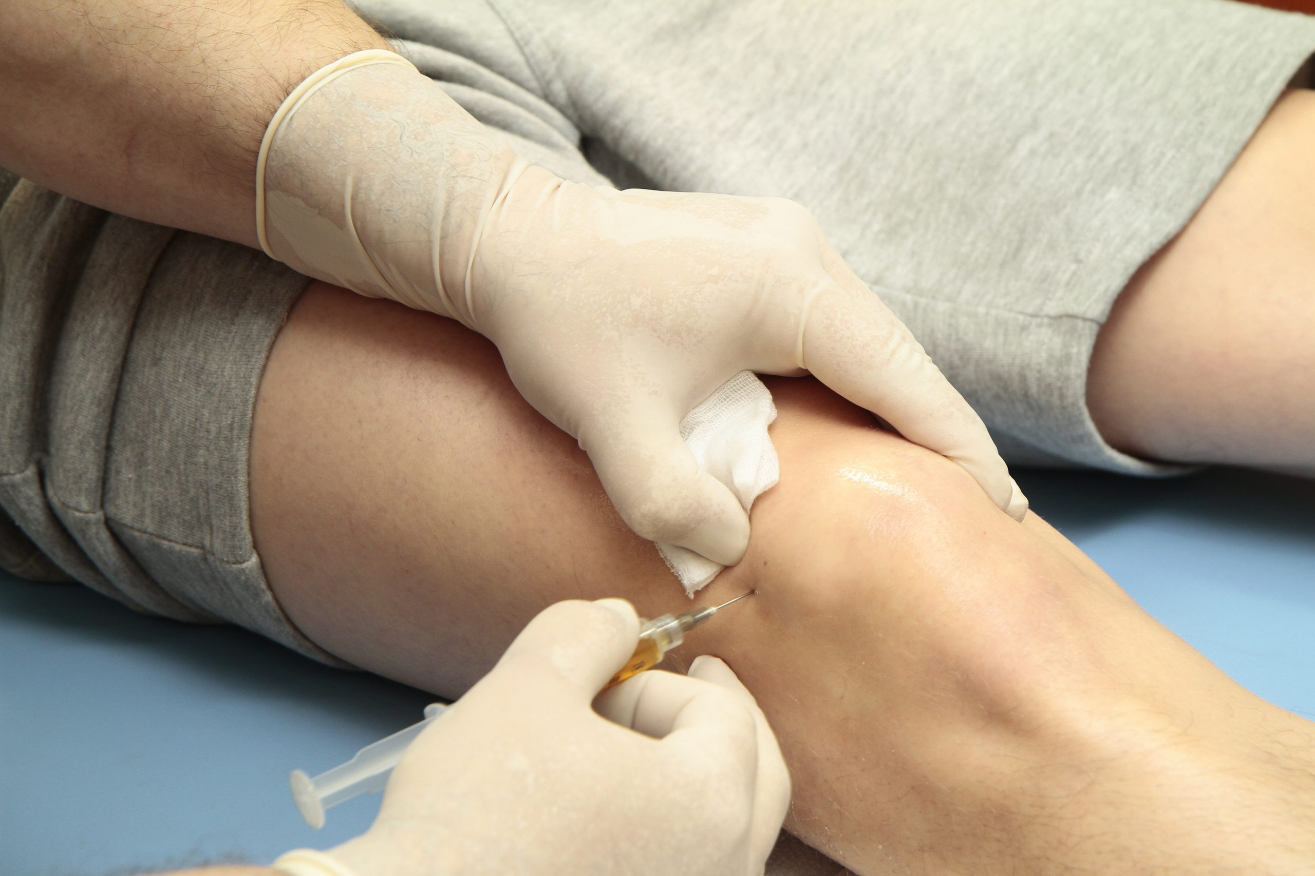 Knees
Knees Hip
Hip Shoulder
ChondroFiller Procedure for Modern Cartilage Repair
04/03/26


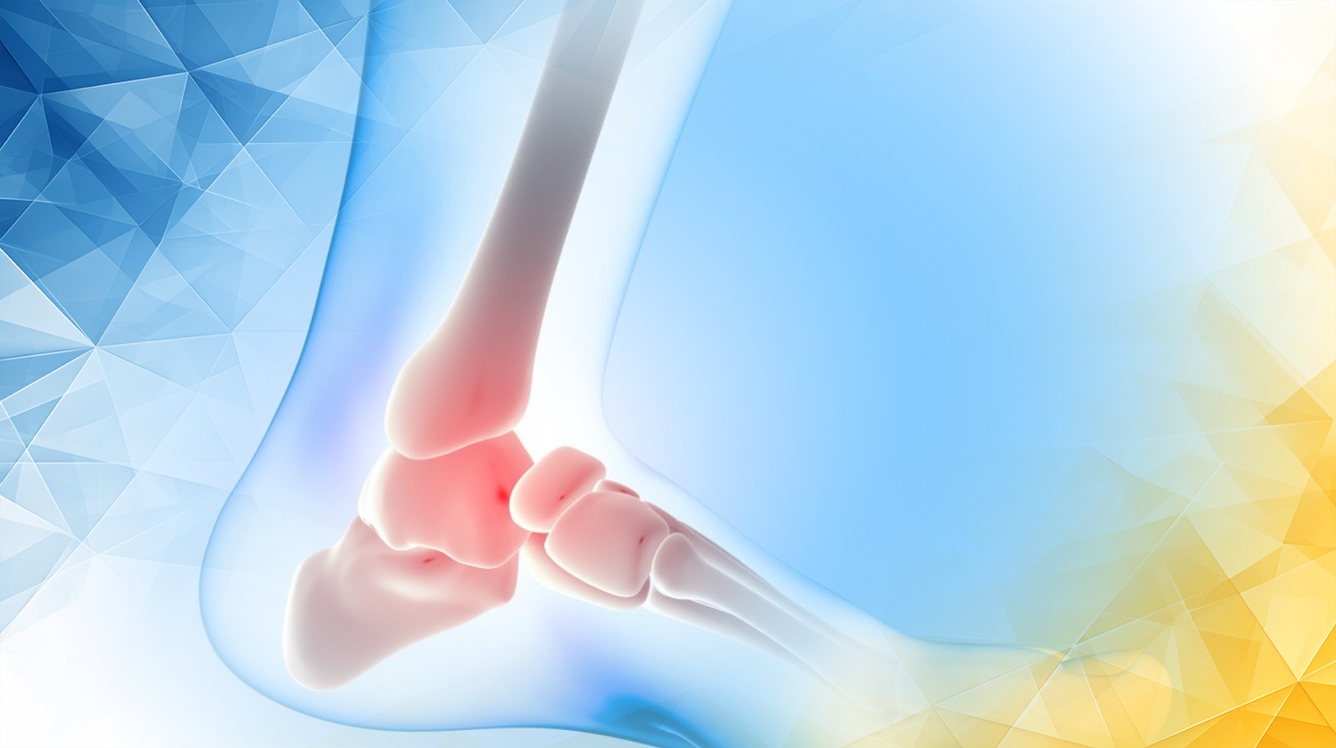
Osteochondritis dissecans (OCD) is a joint condition that often affects the knee , where a small area of cartilage and the underlying bone begin to separate from the rest of the joint surface. What makes OCD tricky is its subtle start: early symptoms are usually mild — perhaps just a bit of discomfort or occasional swelling. Because of this, many people may not realize there’s a problem until it becomes more serious. Without intervention, OCD can progress, leading to significant joint damage, ongoing pain, and even early arthritis that affects both mobility and quality of life.
OCD tends to appear most frequently in adolescents and young adults, though it can affect anyone. Its causes can range from trauma to repetitive stress, or it may arise without a clear reason. Early identification is crucial: the sooner the condition is discovered, the better the chances of protecting the joint and avoiding long-term problems.
Fortunately, advances in medical imaging now make it possible to see tiny, early changes in the knee’s cartilage—before major damage occurs. Thanks to these new technologies, doctors are able to diagnose OCD earlier, choose more effective treatments, and help patients achieve the best possible outcomes.
For many years, standard X-rays have been the main tool for diagnosing OCD. X-rays are good at showing bones, but their big limitation is that they can’t capture problems with soft tissues like cartilage . Early changes in OCD often take place beneath the surface or involve the cartilage , making them invisible on regular X-rays.
This means OCD can go undetected until it’s more advanced and harder to treat. That’s why more sensitive imaging technologies, especially magnetic resonance imaging (MRI), have become essential for catching OCD early and guiding decisions about care.
Early detection really does make a difference. Conventional X-rays can appear normal in the earliest stages, which can make timely diagnosis difficult. MRI bridges this gap by revealing issues before they become obvious on traditional scans, ensuring patients don’t miss out on the opportunity for easier, more effective treatment .
MRI is now considered the gold standard for assessing OCD in the knee. Unlike X-rays, MRI uses magnetic fields and radio waves to provide highly detailed images of both bones and soft tissues—including cartilage .
With specialized MRI techniques, such as T2 mapping, doctors can actually see the microscopic quality and structure of cartilage . This allows them to detect subtle, early changes—like minor cracks or wear long before symptoms become severe or the injury progresses.
These precise images help clinicians determine whether an OCD lesion is stable or unstable—a crucial factor in choosing the right treatment option. The latest generation of imaging tests continues to improve, offering even greater detail and allowing for more customized care.
In short, advanced imaging doesn’t just confirm that OCD is present. It helps pinpoint how serious the problem is, which parts of the joint are affected, and what kind of treatment will likely work best.
Healthy knee cartilage acts like a cushion, absorbing shock and ensuring smooth joint movement . But it’s delicate—small abnormalities at the microscopic level can weaken the cartilage and increase the risk of further joint damage .
Thanks to advanced imaging, doctors can spot these micro-mechanical changes early. If the cartilage is only mildly affected, non-surgical treatments, such as physical therapy , rest, and activity modification, may be enough for the knee to recover. However, when imaging shows more significant damage or that the lesion is unstable, surgery might be recommended to repair or stabilize the area.
Being able to see these subtle changes allows clinicians to tailor their approach to each patient, optimizing outcomes and possibly preventing the need for more invasive treatments later. The stage at which OCD is discovered really does impact the prognosis, reinforcing how important early detection has become.
With earlier and more accurate diagnosis through advanced imaging, doctors are increasingly able to recommend conservative treatments for many patients with OCD. When the lesion is stable and cartilage damage is minimal, non-invasive measures like physiotherapy, bracing, or adjusting daily activities can yield excellent results.
Research shows that early intervention with these less aggressive methods often stops OCD from getting worse, eases symptoms, and even eliminates the need for surgery. The aim is to preserve as much of the knee ’s natural cartilage as possible and reduce the long-term risk of arthritis .
The most important factor is timing: the earlier OCD is detected, the better the outlook with conservative care. Advanced imaging has made finding these issues at their earliest, most treatable stage much more achievable.
Imaging technology is advancing faster than ever. Ultra-high-field MRI scanners and cutting-edge computer models are already being studied and offer the promise of even clearer, more detailed pictures of joint health .
These new tools could lead to earlier, more accurate diagnosis and more personalized treatment plans, ensuring each patient receives the care that’s right for them. While these innovations are still being tested, their potential to further improve OCD management is highly promising.
Advanced imaging has transformed how osteochondritis dissecans is diagnosed and treated, making it possible to spot early, subtle changes in knee cartilage that might otherwise go unnoticed. Acting early allows doctors to use conservative treatments that protect the joint and prevent further damage.
By identifying OCD sooner and personalizing care, patients are more likely to avoid surgery and maintain healthy, pain-free knees for years to come. As technology continues to evolve, the role of advanced imaging in helping people with OCD enjoy an active life is only set to grow.
Hashim, S., Morgan, C., & Sarraf, K. M. (2023). Osteochondritis dissecans. British Journal of Hospital Medicine, 84(4), 1-7. https://doi.org/10.12968/hmed.2023.0044
Linden, B. C., Jonsson, K., & Redlund-Johnell, I. (2003). Osteochondritis dissecans of the hip. Acta Radiologica, 44(1), 67-71. https://doi.org/10.1034/j.1600-0455.2003.00020.x
Powell, J. H., & Whipple, T. L. (1986). Osteochondritis dissecans of the talus. Foot & Ankle, 6(6), 309-310. https://doi.org/10.1177/107110078600600606
London Cartilage Clinic offers state-of-the-art advanced imaging and personalised care for OCD. Led by Prof Lee, who has extensive experience in cartilage repair and knee disorders, the clinic delivers early diagnosis and tailored treatments. Their expertise ensures the best chance of recovery and long-term joint health for patients.
Early diagnosis of knee OCD is crucial for preventing serious joint damage and long-term complications like arthritis. With early intervention, patients benefit from conservative treatments, reduced pain, and improved mobility. London Cartilage Clinic excels in using advanced imaging to detect OCD at its earliest, most treatable stage.
Advanced MRI, especially T2 mapping, allows the clinic’s specialists to detect subtle cartilage changes before symptoms worsen. This precise approach guides optimal treatment choices, whether conservative or surgical. At London Cartilage Clinic, patients gain access to the latest imaging, ensuring faster recovery and better preservation of knee function.
For early and stable cases of OCD, the clinic offers physiotherapy, bracing, rest, and activity modification. These conservative options are designed to encourage natural healing, avoid surgery, and protect the joint. Prof Lee and the team create tailored plans to maximise results and support a pain-free return to activity.
Prof Lee is renowned for his advanced surgical skill in cartilage repair and knee preservation. He uses the latest minimally invasive techniques when surgery is necessary, aiming for faster healing and improved long-term outcomes. Patients benefit from Prof Lee’s vast experience and commitment to delivering the highest standard of care.
All our treatments are selected to help patients achieve the best possible outcomes and return to the quality of life they deserve. Get in touch if you have any questions.
At London Cartilage Clinic, we are constantly staying up-to-date on the latest treatment options for knee injuries and ongoing knee health issues. As a result, our patients have access to the best equipment, techniques, and expertise in the field, whether it’s for cartilage repair, regeneration, or replacement.
For the best in patient care and cartilage knowledge, contact London Cartilage Clinic today.
At London Cartilage Clinic, our team has spent years gaining an in-depth understanding of human biology and the skills necessary to provide a wide range of cartilage treatments. It’s our mission to administer comprehensive care through innovative solutions targeted at key areas, including cartilage injuries. During an initial consultation, one of our medical professionals will establish which path forward is best for you.
Contact us if you have any questions about the various treatment methods on offer.
Legal & Medical Disclaimer
This article is written by an independent contributor and reflects their own views and experience, not necessarily those of londoncartilage.com. It is provided for general information and education only and does not constitute medical advice, diagnosis, or treatment.
Always seek personalised advice from a qualified healthcare professional before making decisions about your health. londoncartilage.com accepts no responsibility for errors, omissions, third-party content, or any loss, damage, or injury arising from reliance on this material. If you believe this article contains inaccurate or infringing content, please contact us at [email protected].