 Cartilage
 Knees
Knees Hip
Hip Shoulder
Ankle Cartilage Damage Causes Symptoms and Latest Treatments
24/02/26
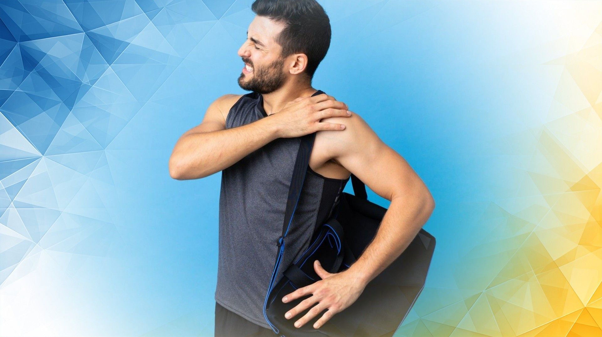

Shoulder instability is a common problem, often caused by injuries such as Hill-Sachs deformity and labrum tears. Simply put, a Hill-Sachs deformity is a dent or groove that forms on the ball of the shoulder joint (the humeral head) when it forcefully strikes the edge of the socket during a dislocation . The labrum, a ring of cartilage, acts as a stabilizer by deepening the shoulder socket. When this cartilage tears —a condition called a labrum tear—the joint becomes more prone to instability.
When these two injuries occur together, the shoulder can become especially unstable and more challenging to treat. In this article, we’ll break down how these injuries happen, explore how doctors use imaging to diagnose them, and review the latest treatment options. We’ll also clarify terms like “Hill-Sachs deformity radiology” and “Bankart lesion vs Hill-Sachs lesion” in straightforward language, drawing on recent medical research. Let’s start by looking at how these injuries develop.
Picture your shoulder joint as a golf ball (the humeral head) sitting on a tee (the socket). During a shoulder dislocation , the ball can slam into the edge of the socket, leaving a dent in its surface—a Hill-Sachs deformity. This dent, sometimes called a cortical depression , can grow larger with repeated dislocations, making it even easier for the shoulder to become unstable again.
At the same time, the labrum—the rubbery ring of cartilage around the socket—may be stretched or torn. Think of it as the gasket that provides a tight seal and stability. When it’s torn, the injury is known as a Bankart lesion, and the shoulder loses important support.
The extent of the dent in the humeral head and the degree of labrum tearing both influence how unstable the shoulder becomes. Larger bone dents and more significant tears raise the risk of repeated dislocations. Studies have shown that identifying and understanding these injuries are essential, as they often occur together and require tailored treatment.
To determine the presence and severity of a Hill-Sachs deformity or labrum tear, doctors rely on advanced imaging. Magnetic resonance imaging (MRI) is excellent for viewing soft tissues like the labrum and is used to spot Bankart lesions. Computed tomography (CT) scans provide a detailed look at the bony dent in the humeral head characteristic of a Hill-Sachs deformity. Sometimes, if the dent is located at the front of the humeral head, it’s called a “reverse Hill-Sachs lesion.”
Ultrasound can sometimes reveal these injuries , though it’s less commonly used. When people refer to “Hill-Sachs deformity radiology,” they’re talking about the range of imaging methods—especially MRI and CT—that help measure the injury and guide treatment decisions. Research increasingly shows that combining MRI and CT scans offers the most complete assessment, letting doctors see both the bone and cartilage injuries and plan the best path forward.
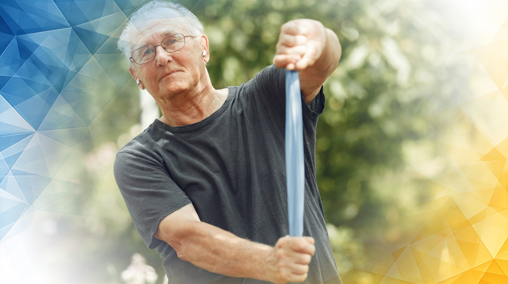
The best way to treat a Hill-Sachs deformity with a labrum tear depends on how severe the injuries are and on the patient’s activity level and health. Sometimes, physical therapy focused on strengthening the shoulder can improve stability and help avoid surgery. This approach is often used for milder injuries or in less active patients.
However, if the dent in the bone is large or the labrum tear is significant, surgery may be recommended. A common minimally invasive option is arthroscopic remplissage, which fills in the dent with soft tissue so it doesn’t catch on the socket and cause more dislocations . Another surgical technique, known as acute Hill-Sachs reduction, aims to restore the normal shape of the humeral head to improve joint function.
These advanced techniques offer many advantages, including smaller incisions and quicker recovery times compared to traditional open surgery. The choice of treatment ultimately depends on the size and severity of the injuries, the patient’s lifestyle, and the overall stability of the shoulder. Research shows that these newer surgical options can significantly reduce the risk of the shoulder dislocating again and help patients return to their daily activities sooner.
When a Hill-Sachs deformity and a labrum tear occur together, they can create a complex challenge that requires a thoughtful, individualized approach. We’ve explored how these injuries develop, how modern imaging helps diagnose them, and the range of treatments available—from physical therapy to cutting-edge, minimally invasive surgery.
Today, the combination of accurate imaging and personalized surgical techniques offers patients the best chance for full recovery and lasting shoulder stability. Ongoing research is likely to improve both diagnosis and treatment options even further in the years ahead. With these advances, more people can regain their shoulder strength and mobility—and get back to the activities they love.
Jeong, J. Y., Stead, T. S., Kwon, J., Carman, M., & Ganti, L. (2022). Hill-Sachs Deformity. Orthopedic Reviews, 14(1). https://doi.org/10.52965/001c.31927
Oh, J.-R., Song, H.-C., Park, J.-G., & Kim, M.-S. (2014). Fused 99mTc-HDP SPECT/MR Imaging of Reverse Hill-Sachs Deformity. Clinical Nuclear Medicine, 39(1), e99–e100. https://doi.org/10.1097/rlu.0b013e3182816339
Solomon, L. Z., Goldwasser, B., Huston, K., & Meltzer, J. A. (2021). Utility of point-of-care ultrasound for the diagnosis of Hill-Sachs deformity in the pediatric emergency department. Pediatric Emergency Care, 37(1), 41-43. https://doi.org/10.1097/pec.0000000000002086
All our treatments are selected to help patients achieve the best possible outcomes and return to the quality of life they deserve. Get in touch if you have any questions.
At London Cartilage Clinic, we are constantly staying up-to-date on the latest treatment options for knee injuries and ongoing knee health issues. As a result, our patients have access to the best equipment, techniques, and expertise in the field, whether it’s for cartilage repair, regeneration, or replacement.
For the best in patient care and cartilage knowledge, contact London Cartilage Clinic today.
At London Cartilage Clinic, our team has spent years gaining an in-depth understanding of human biology and the skills necessary to provide a wide range of cartilage treatments. It’s our mission to administer comprehensive care through innovative solutions targeted at key areas, including cartilage injuries. During an initial consultation, one of our medical professionals will establish which path forward is best for you.
Contact us if you have any questions about the various treatment methods on offer.
Legal & Medical Disclaimer
This article is written by an independent contributor and reflects their own views and experience, not necessarily those of londoncartilage.com. It is provided for general information and education only and does not constitute medical advice, diagnosis, or treatment.
Always seek personalised advice from a qualified healthcare professional before making decisions about your health. londoncartilage.com accepts no responsibility for errors, omissions, third-party content, or any loss, damage, or injury arising from reliance on this material. If you believe this article contains inaccurate or infringing content, please contact us at [email protected].